・Letter to the Editor・・Current Issue・ ・Achieve・ ・Search Articles・ ・Online Submission・ ・About IJO・
Hydration with
Cefuroxime-a method for sealing a small leaking corneal perforation
Gilad Allon, Itzchak Beiran, Eytan Z.
Blumenthal
Department of Ophthalmology, Rambam Health Care Campus, Haifa
3109601,
Israel
Correspondence to: Eytan Z. Blumenthal.
Department of
Ophthalmology, Rambam Health Care Campus, Haifa 3109601, Israel.
e_blumenthal@rambam.health.gov.il
Received:
2014-11-02
Accepted: 2015-03-04
DOI:10.18240/ijo.2016.05.27
Citation: Allon
G, Beiran I, Blumenthal EZ. Hydration with
Cefuroxime-a method for sealing a small leaking corneal perforation. Int J Ophthalmol 2016;9(5):792-793
Dear Sir,
I am Dr. Gilad Allon from the Department of Ophthalmology of the
Rambam Health Care Campus, Haifa, Israel. I write to present a case report of
corneal hydration with Cefuroxime in order to seal a traumatic corneal
perforation.
In the case of a penetrating trauma limited to the cornea, the immediate
objective of treatment is to seal the globe and avoid later intraocular
infection. Sealing the globe can be achieved by suturing, glueing with tissue
adhesives or applying a soft contact lens together with an aqueous suppressant.
Suturing is mandatory for larger and irregular perforations, while glueing and
a contact lens may be preferred in cases of small corneal perforations[1-3]. In such cases often the
sole antibiotic coverage includes a topical antibiotic[4-6].
A healthy 29 year-old man was admitted to the emergency room with a small
(1.5 mm long) central (1 mm off axis) linear perforation
occurring 2h prior to admission. The anterior chamber was almost flat, and a
constant leak noted. Visual acuity was finger counting from 3 meters. The
trauma was caused by a metallic wire fence. No iris or lens damage was noted,
and no intraocular foreign body was identified. Owing to the small and central
location, it was decided not to suture the opening. A soft therapeutic contact
lens was placed on the eye, deepening the chamber within an hour. The patient
was hospitalized and placed on an aqueous suppressant, a topical wide-spectrum
antibiotic as well as on a systemic antibiotic. Upon daily inspection the
anterior chamber remained deep and relatively quiet, until 6d after admission,
the contact lens was removed to check for any remaining leak. Unfortunately a
steady leak was present (Figure 1).
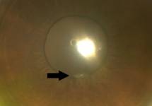
Figure 1 The
perforation site before hydration.
After considering the various options, stromal hydration was performed at
the slit-lamp in a sterile fashion, after application of povidone iodine. The
procedure was performed using a 27 G cannula on a 1
mL syringe
filled with 0.3 mL Cefuroxime at a concentration of 1
mg/0.1
mL.
The technique
applied is identical to the approach used to seal leaking incisions at the end
of cataract surgery. In addition, some of the antibiotic was intentionally
squirted into the anterior chamber. Immediately following the stromal hydration
the leak stopped and did not recur at any time following the procedure (Figure
2). A month following the injury visual acuity was 0.9 and the anterior segment
was normal short of a small full-thickness linear corneal scar.
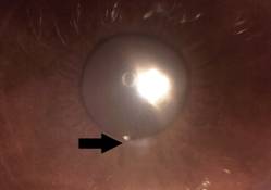
Figure 2 The perforation
site after hydration.
We described an unusual approach to sealing a small corneal perforation,
using a method routinely used to seal small "perforations" inflicted
on the eye during routine cataract surgery. Instead of balanced salt solution,
we chose to use Cefuroxime, the antibiotic of choice we inject at the end of
each cataract surgery performed at our institute[6-8]. Cefuroxime injected into
the corneal stroma was previously described as a treatment for infectious
crystalline keratopathy[9].
While we performed this procedure only 6d post-injury, on hind sight it
would have been wiser to perform it much earlier, ideally when the patient was
first seen. This could have led to closure of the leak, as well as delivered an
antibiotic into the anterior chamber, soon after the suspected contamination,
as opposed to 6d later.
ACKNOWLEDGEMENTS
Conflicts
of Interest: Allon G, None;
Beiran I, None;
Blumenthal EZ, None.
REFERENCES
1 Chan SM,
Boisjoly H. Advances in the use of adhesives in ophthalmology. Curr Opin
Ophthalmol 2004;15(4):305-310. [CrossRef] [PubMed]
2 Castiblanco CP,
Adelman RA. Sympathetic ophthalmia. Graefes Arch Clin Exp Ophthalmol
2009;247(3):289-302. [CrossRef]
[PubMed]
3 Madhusudhan AP,
Evelyn-Tai LM, Zamri N, Adil H, Wan-Hazabbah WH. Open globe injury in Hospital
Universiti Sains Malaysia - A 10-year review. Int J Ophthalmol 2014;7(3):486-490.
[PMC free article]
[PubMed]
4 Cooper BA,
Holekamp NM, Bohigian G, Thompson PA. Case-control study of endophthalmitis
after cataract surgery comparing scleral tunnel and clear corneal wounds. Am J
Ophthalmol 2003;136(2):300-305. [CrossRef]
5 Miller JJ, Scott
IU, Flynn HW Jr, Smiddy WE, Newton J, Miller D. Acute-onset endophthalmitis
after cataract surgery (2000-2004): incidence, clinical settings and visual
acuity outcomes after treatment. Am J Ophthalmol 2005;139(6):983-987. [CrossRef] [PubMed]
6 Seal DV, Barry
P, Gettinby G, Lees F, Peterson M, Revie CW, Wilhelmus KR; ESCRS
Endophthalmitis Study Group. ESCRS study of prophylaxis of postoperative
endophthalmitis after cataract surgery: case for European multicenter study. Cataract
Refract Surg 2006;32(3):396-406. [CrossRef] [PubMed]
7 Ataş M, Başkan
B, Ozköse A, Mutlu Sarıgüzel F, Demircan S, Pangal E. Effects of moxifloxacin
exposure on the conjunctival flora and antibiotic resistance profile following
repeated intravitreal injections. Int J Ophthalmol 2014;7(5):855-859.
8 Kocak I, Kocak
F, Teker B, Aydin A, Kaya F, Baybora H. Evaluation of bacterial contamination
rate of the anterior chamber during phacoemulsification surgery using an
automated microbial detection system. Int J Ophthalmol 2014;7(4):686-688. [PMC free article]
[PubMed]
9 Khan IJ, Hamada
S, Rauz S. Infectious crystalline keratopathy treated with intrastromal
antibiotics. Cornea 2010;29(10):1186-1188.<bb> [CrossRef] [PubMed]
[Top]