·Basic
Research··Current Issue· ·Achieve· ·Search
Articles· ·Online
Submission· ·About IJO· PMC
Inhibiting the effect of 90Sr-90Y ophthalmic
applicators on rat corneal neovascularization induced by sutures
Hong-Yan Zhou1, Shuang Wang1,
Hong Zhang1,Ling Wang1, Wen-Song Zhang 2
1Department of Ophthalmology, China-Japan Union
Hospital of Jilin University, Changchun 130033, Jilin Province, China
2Department of Ophthalmology, the Second Hospital
of Jilin University, Changchun 130033, Jilin Province, China
Correspondence to: Wen-Song
Zhang.
Department of Ophthalmology, the Second Hospital of Jilin University, Changchun
130033, Jilin Province, China. zhangzhou89@sina.com
Received: 2016-01-21 Accepted:
2016-04-11
Abstract
AIM:
To investigate a practical technique used to inhibit
corneal angiogenesis with a 90Sr-90Y ophthalmic applicator.
METHODS:
A 90Sr-90Y ophthalmic applicator
was detected with a radioactive nuclide application treatment healthy
protection standard. The applicator used was produced through medical dosimetry
research; it had a concave applicator add measured the applicator temperature, serviceable humidity range, applicator
appearance status, applicator
radiation homogeneity, radioautography, and
radiological safety of the
original applicator surface. A vessel model was
established using newborn rats, with sutures around the
corneal limbus. Corneal neovascularization
(CNV) were observed with a slit lamp. The new vessel length and response
area were measured.
RESULTS: Low-dose
radiation can inhibit CNV after corneal sutures. The absorbed dose of the
applicator (0.046 Gy/s) was safe for the treatment of it. The lengths of new
vessels and the areas of new vessels were lower than the new born vessel rat
group (P<0.01).
CONCLUSION:
The optimal radiation dose emitting from the
applicator can be safe and potentially used in humans.
KEYWORDS: radiation; cornea;
neovascularization.
Citation: Zhou HY,
Wang S, Zhang H, Wang L, Zhang WS. Inhibiting the effect of 90Sr-90Y
ophthalmic applicators on rat corneal neovascularization induced by sutures. Int J Ophthalmol 2016;9(9):1251-1254
INTRODUCTION
Corneal neovascularization (CNV) leads
to decreased corneal transparency, which can induce vision impairment and even lead to blindness. The complex process of CNV
is related to multiple factors. Angiogenesis is a highly organized
sequence of cellular events stemming from vascular initiation, formation,
maturation, remodelling and regression processes controlling and modulating
tissue requirements[1].
Pathological CNV caused by infectious, traumatic and degenerative diseases
depends on the imbalance of angiogenic and anti-angiogenic factors[2]. Although many studies have described how to treat newborn corneal
vessels, no exact method has been detailed until now. New therapeutic
strategies must be explored. 90Sr/90Y
is a key example of a high-beta-energy-emitting radionuclide that is available
from the strontium-90 (90Sr)/90Y radionuclide generator
system[3-5]. Using such electrochemical techniques, the lower consumption of reagents
and minimal generation of radioactive waste are compatible with Ą°green
chemistryĄą principles[6].
Radium beta emitters involving 90Sr/90Y applicators have been widely used to treat
superficial skin haemangioma in early childhood[7].
This study aimed to investigate the
effects of brachytherapy
with low-dose beta radiation emitted by 90Sr/90Y on CNV
in a model using sutures and newborn rat vessels.
MATERIALS AND METHODS
90Sr-90Y Ophthalmic Applicator Design The applicator used in this experiment is routinely used
in clinical nuclear medicine. It was purchased from the Atomic Energy Isotope
Research Institute (China), and it was detected using radioactive nuclide application treatment healthy
protection standards. The applicator
that was used was produced through medical dosimetry research; it had a concave
applicator and measured the applicator temperature, serviceable humidity range,
applicator appearance status, applicator radiation homogeneity, radioautography, and radiological
safety of the original applicator surface.
Newborn Rat Vessel Model with
Sutures This study
followed the guidelines of the Guide for the Care and Use of Laboratory
Animals, as well as the principles of the ARVO Statement for the Use of Animals
in Ophthalmic and Vision Research. Two-month-old female Wistar rats without eye
diseases (Animal Experiment Department, Jilin University, China) weighing
200-250 g were anesthetized with a 1% pentobarbital sodium intraperitoneal
injection (1.3 mg/kg). We established a
newborn rat vessel model with sutures (34 rats). Three 10/0
nylon sutures were applied to the limbus of the cornea.
Observation
and Examination The
54 newborn rats were randomly divided into three groups: the control group
(radiation treatment without sutures, n=17),
the suture group (no radiation treatment, n=17)
and the irradiation treatment with sutures group (IT, n=17). Rats in the IT and control groups received a low dose of 7
Gy once daily for 7 consecutive days. All rats in each group were clinically
evaluated. At the end of the experiment, all rats were sacrificed with an
overdose of 1% pentobarbital sodium. The corneas of the rats were harvested,
and only the right eye of each rat was used. We randomly selected six rats from
each group, and we observed the neovascularization
using a slit lamp. We calculated the new vessel length and response area using the Needleman et al[8] method. The data were statistically analysed.
Measurement of the Applicator
A SSR9013
applicator (Figure 1) was
used; the absorbed dose rate was 0.046
Gy/s, and the absorbing dose rate was 0.0345 Gy/s. The applicator measurement
followed the radioactive nuclide application
treatment for health protection standard.

Figure 1 An 90Sr-90Y ophthalmic applicator This SSR9013 applicator was consistent with the radioactive nuclide application treatment health protection standard.
Manufacture research: the spherical
source was made by nuclide 90Sr-90Y. The applicator was
fit to the corneal spherical surface. Medical dosimetry research: the dosimetric measurement and spatial
distribution of the applicator were useful for determining clinical needs. The
applicator concave surface contamination level was less than 185 Bq. The applicator temperature was 5Ąæ-40Ąæ, and the serviceable humidity range was less than 80%. In terms of the
applicator appearance status, the
applicator had an integrated surface, and no radioactive substance was divulged. Radioautography:
the radioactive source surface of the ophthalmic applicator was placed above
the sensitization film and then removed after 1 to 5s. The analysis of the
printed film showed the homogeneity of
the applicator (Figure 2).
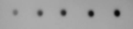
Figure 2 Radioautography The radioactive source surface of the ophthalmic
applicator was placed above the sensitization film and then removed after 1 to
5s. The ophthalmic
applicatorĄŊs autoradiography results
showed homogeneity.
The radiant security of the applicator: the
maximum beta radiation level emitted by the 90Sr/90Y
applicator was 0.546 MeV. The range of 0.546 MeV in the tissue was 0.246 cm.
The distance between the applicator and the cornea was 1 mm, and the central
corneal thickness was 0.6 mm. The average anterior chamber depth was 0.27 mm.
Therefore, the dosage used in this experiment was safe enough to eliminate
cataracts and relative complications.
Statistical Analysis The lengths
and areas of the new vessels were analysed, followed by one-factor analysis of
variance to compare the between-group differences, and P<0.05 was considered to be statistically significant.
RESULTS
Measurement
of New Vessel Length for Corneal Neovascularization Using the Needleman et al[8] method, six
rats were randomly selected from each group on the 7th day. The
average new vessel length (VL) was calculated as follows. The cornea was
divided into four quadrants. The values of the longest vessel from the four
quadrants were summed, and the average equalled the VL value. The IT group
manifested a VL value that was significantly lower than that of the newborn rat
vessel model group. The statistical analysis was followed by one-factor
analysis of variance, and P<0.01
was considered to be statistically significant compared to the newborn rat
vessel model group (Figure 3).

Figure
3 The IT group manifested a VL value that was significantly lower than that of
the newborn rat vessel model group bP<0.01 was considered to be
statistically significant compared to the newborn rat vessel model group.
Measurement
of New Vessel Area for Corneal Neovascularization Following the Needleman et al[8]
method, six rats were randomly selected from each group on the 7th
day. The average new vessel area was calculated using the following formula:
area (mm2)=CH/12ĄÁ3.14[r2-(r-VL)];
r=3 mm; CH measured the hours of new vessels. The value of the new vessel area
in the IT group was much lower than that in the newborn rat vessel model group.
The statistical analysis was followed by one-factor analysis of variance, and P<0.01 was considered to be
statistically significant compared to the newborn rat vessel model group
(Figure 4).

Figure
4 The IT group manifested an average new vessel area that was
significantly lower than that of the newborn rat vessel model group aP<0.05 was considered to be
statistically significant compared to the newborn rat vessel model group.
The CNV length and area were much lower
in the IT group compared with the newborn rat vessel model on the seventh day
after the experiment (length: P<0.01; area: P<0.01). The statistical analysis was followed by
one-factor analysis of variance, and P<0.01
was considered to be statistically significant compared to the newborn rat
vessel model group (Figures 3, 4).
Western Blot Analysis Proteins were
extracted from
the rat corneas on the 7th day after suturing. Equal amounts
of proteins extracted from lysates were subjected to electrophoresis on 10%
Tricine gels and then electrophoretically transferred to PVDF membranes. After
1h of blocking in 0.05
g/mL milk, the blots were incubated with primary antibodies against vascular endothelial growth factor (VEGF) at 4ĄãC
overnight. After washing three times with Tris-buffered saline with 0.05% Tween
20 for 10min each, the membranes were incubated with horseradish Peroxidase (HPR)-conjugated goat
anti-rabbit IgG for 1h at room temperature. The specific bands were visualized
using enhanced chemiluminescence reagents and recorded on film (Figure 5).

Figure
5 Effect of irradiation on VEGF expression after corneal suturing The Western
blot analysis results showed that VEGF was expressed in normal rat corneas and
that this expression increased from day 3 and peaked at day 7. According to the
data, the VEGF expression was much lower in the irradiation group compared to
the alkali burn group on the 7th day.
DISCUSSION
Angiogenesis, the process by which new blood
vessels arise from pre-existing vessels, is a critical part of many disease
processes, including CNV[9-12]. Regulation of angiogenesis is crucial
for many diseases. Antiangiogenic therapy focusing on the tumour
microenvironment is a traditional approach for treating cancer[13].
Angiogenesis is a complex process that includes multiple cell types, cytokines,
adhesion molecules, growth factors, and signal transduction during inflammation[14]. Radiotherapy for surgery and tumour therapy was developed
more than a century ago. Higher doses of radiotherapy that minimize absorbance
by normal tissues will be the next development trend[15]. Radiation
damage to ocular tissues includes conjunctivitis, eyelid lesions, keratitis,
and keratoconjunctivitis sicca[16]. Appropriate radiation therapy has
long been attempted for ocular diseases. Intraocular tumours located in the
iris, ciliary body, and choroid could be treated
with plaque brachytherapy, except for tumours with orbital extension and no
light perception vision[17].
Endothelial
dysfunction has been associated with a number of pathophysiological processes.
VEGF, which is synthesized and released by endothelial cells, regulates
angiogenesis, vascular tone and permeability[18-20].
The formation of CNV is dependent upon VEGF, as well as the proliferation of
vascular endothelium, remodelling of extracellular matrix components and the
activation of cytokines[21]. VEGF
plays a major role in the process of vessel branch formation, leading to aberrant angiogenic responses. It also plays
important roles in many diseases[22-24]. Radium
applicators and pure beta emitters have been widely used in the past to treat
skin haemangioma in early childhood[25]. High
expression levels of VEGF have been associated with a poor prognosis in cancer
patients, indicating that VEGF could be linked to the efficacy of radiotherapy[26]. VEGF is a valuable molecular
marker in treatment outcomes following radiation therapy for rectal
adenocarcinoma[27]. Radiation
therapy can also provide a dose-dependent benefit in the treatment of
neovascular age-related macular degeneration, which can reduce the frequency of
anti-VEGF injections to maintain visual acuity[28].
The
side effects of radiotherapy prevent widespread radiotherapy usage in ocular
disease. In this study, we investigated the potential role of low-dose radiation
for CNV therapy. CNV is a major cause of blindness and can lead to keratoplasty
failure. We focused on the available therapeutic options for CNV. The effect of
90Sr-90Y ophthalmic applicator on CNV was unknown. Based
on the data from our investigation, low doses of beta emissions divided several
times showed good outcomes. The inhibition effect peaked on the 7th day after irradiation
therapy. The irradiation distance is safe; it cannot induce cataract and
corneal opacity. There were significant differences in the average new vessel
length and new vessel area in the suturing group and the IT group, as we
predicted. We made these observations using lamp light, and we found that
divided low-dose radiation performed better than high-dose radiation or therapy
only once (data not shown). The corneas were transparent, without vessels. The
vessels terminated at the corneal limbus. Angiogenesis occurs in corneal
pathologies and wounds, and new vessels leak easily. Exudation and fibrosis of
new vessels can lead to blindness. It is hard to treat CNV. Until now, there
has been no definitive approach. Radiation can inhibit tumour
neovascularization, particularly in neonatal angioma. A suitable dose of
radiation can be safe enough to inhibit the CNV induced by sutures. The
therapeutic effect of CNV is unclear.
CNV occurs in corneal injury pathologies, such as infection, chemical
burns, physical trauma, and corneal transplant
rejection[29-30]. The data above clearly suggest that radiation therapy can be used to treat CNV. 90Sr-90Y ophthalmic applicators may provide new insight into
the treatment of angiogenic ocular surface diseases. Therefore, further
studies are needed to determine the appropriate dosage and the precise
mechanism of the inhibiting effect of irradiation. In conclusion, this study
demonstrates that radiation may be a new pragmatic method to treat corneal
vessels. Thus, we conclude that 90Sr-90Y ophthalmic applicators have the
potential to become an ideal platform for CNV therapy.
ACKNOWLEDGEMENTS
Foundations: Supported by National Natural Science Foundation (No.81300727);
the Research Fund of Jilin Provincial Science and Technology Department
(No.20160101011JC).
Conflicts of Interest: Zhou HY, None; Wang S,
None; Zhang H, None; Wang L, None; Zhang WS,
None.
REFERENCES
1 Staton CA, Reed MW, Brown NJ. A critical analysis of
current in vitro and in vivo angiogenesis assays. Int J Exp Pathol 2009;90(3):195-221. [CrossRef] [PubMed] [PMC free article]
2 Chang JH, Gabison EE, Kato T, Azar DT. Corneal
neovascularization. Curr Opin Ophthalmol 2001;12(4):242-249. [CrossRef]
3 Montaña RL, GonzĻĒlez IH, Ramirez AA, Garaboldi L,
Chinol M. Yttrium-90-current status, expected availability and applications of
a high beta energy emitter. Curr
Radiopharm 2012;5(3):253-263. [CrossRef]
4 Saghamanesh S, Karimian A, Abdi M. Absorbed dose
assessment of cardiac and other tissues around the cardiovascular system in
brachytherapy with 90Sr/90Y source by Monte Carlo simulation. Radiat Prot Dosimetry 2011;147(1-2):296-299.
[CrossRef] [PubMed]
5 Schiele TM, Herbst J, Pöllinger B, Rieber J, König
A, Sohn HY, Krötz F, Leibig M, Belka C, Klauss V. Late and very late catch-up
after 90Sr/90Y beta-irradiation for the treatment of coronary in-stent
restenosis. Acute Card Care
2011;13(1):9-13. [CrossRef] [PubMed]
6 Chakravarty R, Dash A, Pillai MR. Electrochemical
separation is an attractive strategy for development of radionuclide generators
for medical applications. Curr Radiopharm
2012;5(3):271-287. [CrossRef]
7 Shamsaldin A, Lundell M, Diallo I, Ligot L,
Chavaudra J, de Vathaire F. Estimation of the radiation dose from radiotherapy
for skin haemangiomas in childhood: the ICTA software for epidemiology. Phys Med Biol 2000;45(12):3589-3599. [CrossRef]
8 Needleman P, Turk J, Jakschik BA, Morrison AR,
Lefkowith JB. Arachidonic acid metabolism. Annu
Rev Biochem 1986;55:69-102. [CrossRef] [PubMed]
9 Gaengel K, Niaudet C, Hagikura K, et al. The sphingosine-1-phosphate
receptor S1PR1 restricts sprouting angiogenesis by regulating the interplay
between VE-cadherin and VEGFR2. Dev Cell
2012;23(3):587-599. [CrossRef] [PubMed]
10 Kumar B, Gupta SK, Srinivasan BP, Nag TC,
Srivastava S, Saxena R. Hesperetin ameliorates hyperglycemia induced retinal
vasculopathy via anti-angiogenic effects in experimental diabetic rats. Vascul Pharmacol 2012;57(5-6):201-207. [CrossRef] [PubMed]
11 Caccuri F, Giagulli C, Bugatti A, Benetti A,
Alessandri G, Ribatti D, Marsico S, Apostoli P, Slevin MA, Rusnati M, Guzman
CA, Fiorentini S, Caruso A. HIV-1 matrix protein p17 promotes angiogenesis via
chemokine receptors CXCR1 and CXCR2. Proc
Natl Acad Sci U S A 2012;109(36):14580-14585. [CrossRef] [PubMed] [PMC free article]
12 Villalvilla A, Moro M, Arruza L, Redondo S,
FernĻĒndez-Cruz A, FernĻĒndez-Durango R. Circulating endothelial progenitor cells
are reduced in rat oxygen-induced retinopathy despite a retinal SDF-1/CXCR4 and
VEGF proangiogenic response. Life Sci
2012;91(7-8):264-270. [CrossRef] [PubMed]
13 Indraccolo S, Mueller-Klieser W. Potential of
induced metabolic bioluminescence imaging to uncover metabolic effects of
antiangiogenic therapy in tumors. Front
Oncol 2016;6:15. [CrossRef] [PubMed] [PMC free article]
14 Alkim C, Alkim H, Koksal AR, Boga S, Sen I.
Angiogenesis in inflammatory bowel disease. Int
J Inflam 2015;2015:970890. [CrossRef] [PubMed] [PMC free article]
15 Choi WH, Cho J. Evolving clinical cancer
radiotherapy: concerns regarding normal tissue protection and quality
assurance. J Korean Med Sci 2016;31
Suppl 1:S75-S87. [CrossRef] [PubMed] [PMC free article]
16 Pinard CL, Mutsaers AJ, Mayer MN, Woods JP.
Retrospective study and review of ocular radiation side effects following
external-beam Cobalt-60 radiation therapy in 37 dogs and 12 cats. Can Vet J 2012;53(12):1301-1307. [PMC free article] [PubMed]
17 American Brachytherapy Society - Ophthalmic
Oncology Task Force, ABS - OOTF Committee; Simpson ER, Gallie B, Laperrierre N,
et al. The American Brachytherapy
Society consensus guidelines for plaque brachytherapy of uveal melanoma and
retinoblastoma. Brachytherapy
2014;13(1):1-14. [CrossRef] [PubMed]
18 Traub F, Schleicher S, Kirschniak A, Zieker D,
Kupka S, Weinmann M, Königsrainer A, Kratt T. Gene expression analysis in
chronic postradiation proctopathy. Int J
Colorectal Dis 2012;27(7):879-884. [CrossRef] [PubMed]
19 Batycka-Baran A, Paprocka M, Krawczenko A, Kantor
A, Dus D, Szepietowski JC. Reduced number of circulating endothelial progenitor
cells (CD133+/KDR+) in patients with plaque psoriasis. Dermatology 2012;225(1):88-92. [CrossRef] [PubMed]
20 Deng X, Szabo S, Khomenko T, Tolstanova G, Paunovic
B, French SW, Sandor Z. Novel pharmacologic approaches to the prevention and
treatment of ulcerative colitis. Curr
Pharm Des 2013;19(1):17-28 [PubMed]
21 Xiao O, Xie ZL, Lin BW, Yin XF, Pi RB, Zhou SY.
Minocycline inhibits alkali burn-induced corneal neovascularization in mice. PLoS One 2012;7(7):e41858. [CrossRef] [PubMed] [PMC free article]
22 Yang L, Wang X, Zhen S, Zhang S, Kang D, Lin Z.
Aquaporin-4 upregulated expression in glioma tissue is a reaction to
glioma-associated edema induced by vascular endothelial growth factor. Oncol Rep 2012;28(5):1633-1638. [PubMed]
23 Lu J, Yao YY, Dai QM, Ma GS, Zhang SF, Cao L, Ren
LQ, Liu NF. Erythropoietin attenuates cardiac dysfunction by increasing
myocardial angiogenesis and inhibiting interstitial fibrosis in diabetic rats. Cardiovasc Diabetol 2012;11:105. [CrossRef] [PubMed] [PMC free article]
24 Gaengel K, Niaudet C, Hagikura K, et al. The sphingosine-1-phosphate receptor
S1PR1 restricts sprouting angiogenesis by regulating the interplay between
VE-cadherin and VEGFR2. Dev Cell
2012;23(3):587-599. [CrossRef] [PubMed]
25 Shamsaldin A, Lundell M, Diallo I, Ligot L,
Chavaudra J, de Vathaire F. Estimation of the radiation dose from radiotherapy
for skin haemangiomas in childhood: the ICTA software for epidemiology. Phys Med Biol 2000;45(12):3589-3599. [CrossRef]
26 Heravi M, Tomic N, Liang L, Devic S, Holmes J,
Deblois F, Radzioch D, Muanza T. Sorafenib in combination with ionizing
radiation has a greater anti-tumour activity in a breast cancer model. Anticancer Drugs 2012;23(5):525-533. [CrossRef] [PubMed]
27 Kim JW, Kim YB, Choi JJ, Koom WS, Kim H, Kim NK,
Ahn JB, Lee I, Cho JH, Keum KC. Molecular markers predict distant metastases
after adjuvant chemoradiation for rectal cancer. Int J Radiat Oncol Biol Phys 2012;84(5):e577-584. [CrossRef] [PubMed]
28 Kishan AU, Modjtahedi BS, Morse LS, Lee P.
Radiation therapy for neovascular age-related macular degeneration. Int J Radiat Oncol Biol Phys
2013;85(3):583-597. [CrossRef] [PubMed]
29 Chan EC, Van Wijngaarden P, Chan E, Ngo D, Wang JH,
Peshavariya HM, Dusting GJ, Liu GS. NADPH oxidase 2 plays a role in experimental
corneal neovascularization. Clin Sci
(Lond) 2016;130(9):683-696. [CrossRef] [PubMed]
30 Gimenez F, Mulik S, Veiga-Parga T, Bhela S, Rouse
BT. Robo 4 Counteracts Angiogenesis in Herpetic Stromal Keratitis. PLoS One 2015;10(12):e0141925. [CrossRef] [PubMed] [PMC free article]
[Top]