INTRODUCTION
Optical modelling of the human eye is a field with a large variety of models, either new or alterations of oldones[1-2]. There has also been research in trying to create an eye model tofit the statistical data collected from healthy people[3]. In the end there is no model that could be widely used in the visual research field. All of them use almost the same parameters, based on the way that they are designed (e.g. mean parameter values based on population, optimized parameters for specific results etc.). Differences between simple and more complicated designs (with three or four refractive surfaces)exist, dependingon the reason for which they are used. Some models are designed either with a simple crystalline lens, or with grading refractive index or even with an accommodating crystalline lens. Each model has advantages and disadvantages,can simulate different procedures, parameters and metrics of the human eye[1,4].
Nevertheless, there is still need for more eye models in order to cover different areas of research such as ray-tracing of nonsymmetric eye models[5] and models with misaligned optical designs[6]. Further investigation and comparisson studies are held and needed because the natural human eye has a robust optical design which differs between people[7].
In this work three different models were used: the Navarro model[1,8], the Arizona model[2], which is accommodating by using mathematical functions and the Liou-Brennan[9], which is a more anatomically accurate model.
The Navarro model[8], wasfirstly created in 1985 and consists out of four centered aspheric refracting surfaces. Each one of them represents a refracting surface of the human eye: two for the cornea and two for the crystalline lens. It also uses a fl at retinal surface, although there are also versions with spherical retinal surface as well. The main characteristic of this model is that it has a fl at surface as a retina. This makes it suitable for on axis simulations. As rays create an angle in respect to the optical axis the image gets defocused and the coma aberration increases. The other models use a spherical surface for the retina. It is a simple model with rotational symmetry, axial length 24 mm and a total dioptric power of 60.4 D.
The Arizona model[2], is also rotationally symmetric. It has the ability to accommodate in different distances by altering its dioptric power. This is done by use of mathematical functions that change the geometry of the crystalline lens, its refractive index and the anterior chamber depth. In this way it can simulate every particular change of its media during the accommodation process. Its axial length is 24.003 mm but the total dioptric power depends on the accommodation distance.A more anatomically accurate model was created by Liou and Brennan in 1997[9]. Its major difference is that it has a gradient refractive index for the crystalline lens instead of a simple one.It is not rotationally symmetric because of the decentration of the pupil by 0.5 mm nasally. This decentration affects aberrations, particularly coma, as well as curvature offield and vignetting. The equivalent power of this model is 60.35 D and its axial length 23.95 mm.
There are accommodative models in our knowledge such as the Arizona that can accommodate by their own and even simulate the ageing of the eye[10]. There are also computer animated models that show how the parts of the eye move with accommodation and studies about the changes in the crystalline lens with ageing and the mechanisms of accommodation[11-16].In our work the accommodation function in accommodative and non-accommodative models is examined and compared.The Arizona model is able to accommodate by the use of mathematical equations for its media. The other two models were designed to be non-accommodative.Values chosen from the literature were inserted in order to simulate accommodation[17].This comparison study will provide us knowledge on simulation of accommodation with different customized models.
Comparing to previous works mentioned above, in this study we compare one accommodative and two non-accommodative models. The main target is to check if non-accommodative models can provide simulations of accommodation. Moreover,there is a comparison with an accommodative model in order to compare the results between them.
MATERIALS AND METHODS
Ray tracing and optical design software (Zemax, USA) was implemented to design and work with the eye models. The models were chosen because they are mostly used in modelling of vision science research. The common feature of the chosen models is that all of them have aspheric refractive surfaces.On the contrary, they use different parameters for simulating the optical procedure of the human eye. These differences are between the retinal surfaces, optical and visual axis, types of crystalline lenses and values for their optical media.
All models that were used in this study were designed following the works published by their designers[2,8-9]. They have four refractive surfaces, two for the cornea and another two for the crystalline lens. In the Liou-Brennan model the crystalline lens has a grading refractive index and was designed by two different parts, one for the anterior part of the lens and one for the posterior. The refractive indices and the thicknesses of all the media were taken from the original articles.
Accommodation in Human Eye Models All previously described models are designed to be non-accommodative,except from the Arizona. The other two models arefixed to focus at infinity, while the Arizona model can focus at any accommodative demand by using the mathematical algorithmsthat come with it. In order to compare accommodation results between these models, the Navarro and Liou-Brennan models had to accommodate as well.
Four different target distances were used to simulate the accommodation process. These were for infinity, 3, 1 and 0.5 m (0, 0.3, 1 and 2 D accommodative demand in respect).Each chosen value of change corresponds to a specific accommodative demand. The target distances were introduced in the softwarein the “Thickness” box of the “Object” line and the units used were millimeters (mm). For these simulations twofields of incoming rays were used, parallel to the optical axis (0 degrees angle) and not parallel (5 degrees angle). These were introduced in the “Field Data Editor” in the software.The wavelength that was used was 587.6 nm, which is the middle one of the “F, d, C (Visible)” wavelength group in the“Wavelength Data Editor”.
In every distance all models were optimized. That was done because of changing their parameters (radii of curvature of the lens, thicknesses of their media etc. as it will be explained in the following paragraphs) in order to accommodate. The models had to be optimized in order to create a clear image on the retinawith the given parameters and to compare their results. This optimization was done with the “Optimization Tool”of the software. This uses a “merit function” that minimizes the root mean square (RMS) error in the retinal plane and is defined as,![]() where MF is the merit function value, W is the weight factor for each operand used for the calculation of the merit function value, V is the value of each operand, T is the target value for each operand and the factor “i” is for all the population of the operands. The softwaretook into account the variable that was chosen and made all the calculations in order to minimize the merit function value. In the end it returned the best value of the chosen variable.For the optimizations in this study a “Default Merit Function”was implemented to calculate the least RMS wavefront error in the centroid of the image created in the retina. In the “Pupil Integration Method”, 3 rings and 6 arms of incoming rays was used in a “Gaussian Quadrature”.
where MF is the merit function value, W is the weight factor for each operand used for the calculation of the merit function value, V is the value of each operand, T is the target value for each operand and the factor “i” is for all the population of the operands. The softwaretook into account the variable that was chosen and made all the calculations in order to minimize the merit function value. In the end it returned the best value of the chosen variable.For the optimizations in this study a “Default Merit Function”was implemented to calculate the least RMS wavefront error in the centroid of the image created in the retina. In the “Pupil Integration Method”, 3 rings and 6 arms of incoming rays was used in a “Gaussian Quadrature”.
As an optimization variable was selected the vitreous thickness.That was done because this was the only free parameter while the model was getting optimized. During accommodation the corneal parameters (curvatures and thicknesses) and refractive indices of all media do not change. The anterior chamber depth, the crystalline lens thickness and the curvatures of the crystalline lens surfaces are changing and set by us.
While simulating accommodation by changing the target distances and the parameters mentioned above, if the chosen values for the parameters are not suitable, then there will be an obvious change in the total length of the eye. This change will occur through the optimization process which will try to minimize the RMS wavefront error in the retina by changing only the vitreous thickness.
The changes of the ocular system during accommodation are known and studied thoroughly before. All these changes happen with a small increase in the total length of the eye[17-22].In order to make the Navarro and Liou-Brennan models to accommodate, specific values from the literature were used[17].While accommodating the anterior chamber depth decreases and the crystalline lens thickness increases. The crystalline lens changes it’s shape to become more spherical by decreasing it’s radii of curvature. The pupil diameter also decreasein order to increase the depth of focus.
The refractive index of the crystalline lens naturally is gradient.This means that its dioptric power depends on the geometrical characteristics of the lens. There are two ways to simulate this dependency: either to change only the geometrical characteristics (Navarro and Liou-Brennan models) or to change the refractive index of the lens too (Arizona model).
The parameters that were changed during accommodation were: the anterior chamber depth between 3.35 and 3.23 mm,lens thickness between 3.85 and 4.03 mm (corresponding“Thickness” box of each line in the software), anterior radius of curvature of the crystalline lens between 12.8 and 11.5 mm and posterior radius between 5.96 and 5.22 mm (corresponding“Radius” box of each line in the software). The pupil radius also changed from 2 to 1.5 mm (corresponding “Semi-Diameter” box of each line in the software). These changes occur while the target comes closer to the model. All values were chosen as mean values of parameter changes in order to accommodate, following the concept that all eye models are designed with mean values of real human eyes.

Figure 1 Letter F diffraction images In this Figure are shown the images of a letter F as they are simulated in the retinal plane of the eye models in each target distance. The Arizona model is optimized in all distances, as an accommodative model.
The Arizona model has an algorithm that makes it accommodative[2]. In this way is optimized in all distances. The algorithm has mathematical functions that change the geometrical characteristics of the crystalline lens and the aqueous thickness. The refractive index of the crystalline lens in this model also changes while accommodating. This happens because in this model a simple refractive index is used. The physiological refractive index is gradient and while accommodating is dependent on the geometric characteristics of the crystalline lens. In order to simulate this change, this model slightly changes the index while it accommodates.
In this model the same values were used as in the Navarro.The difference here is the procedure of changing the thickness of the lens. In this model the crystalline lens is divided in two parts with different thicknesses and with a grading refractive index. In order to set the crystalline lens thickness, firstly the total thickness of the unaccommodated lens (in 0 D) was subtracted from the needed in each accommodation level. The result was divided by 2, and each half was added to each of the parts. In other words, it was assumed that the two parts change their thickness equally while accommodating.
RESULTS
In the following results “non-optimized” refers to each model coming from the literature, without any optimization. “Optimized”refers to each model after the optimization process that was mentioned earlier.
For graphical comparison, image diffraction analysis was used in the ray tracing and optical design software. In Figure 1 there is a comparison between letter F diffraction images from each model at all distances. As expected, the image gets clearer as the target approaches to the model. Moreover, in Navarro and Liou-Brennan models are obvious the differences between optimized and non-optimized results at infinity.

Figure 2 Differences between optimized and non-optimized models at infinity In spot diagrams the dimension is in μm. In MTF diagrams the dashed line shows the diffraction limit for a pupil radius of 2 mm. The double line in the Liou-Brennan MTF diagrams is because of the decentration of the pupil.
Spot diagrams show the intersection of a ray pattern with the retinal field. Modulation transfer function (MTF) diagrams show the response of the models in each accommodative level,between 0 and 100 cycles/mm.
In Figure 2 the differences between optimized and nonoptimized models at infinity are more obvious in the Navarro model. In both models, optimized spot diagrams are smaller from the non-optimized ones and the MTF diagrams show better results for the middle spatial frequencies.
In Figure 3 are shown the spot diagrams for three different target distances. It is obvious that the diameter decreases as the target approaches the models. In Liou-Brennan model, the decentration of the pupil creates a “tail” in the spot diagrams(coma aberration).
In Figure 4 the MTF diagrams show again better results as the target distance decreases, which is expected. In the Arizona model the results are almost diffraction limited. This model is designed to have the best results as a perfect optical system that simulates a real eye. The diffraction limit decrease in thefigures, as the target distance decrease. That happens because of the decrease of the pupil.
The total eye lengths of the optimized Navarro and Liou-Brennan models are shown in Table 1. It is obvious that both models change their total length while accommodating. The Navarro model shows smaller difference in its width than the Liou-Brennan. These total lengths are different from the ones that were mentioned earlier and that’s because of the optimization method that was applied.
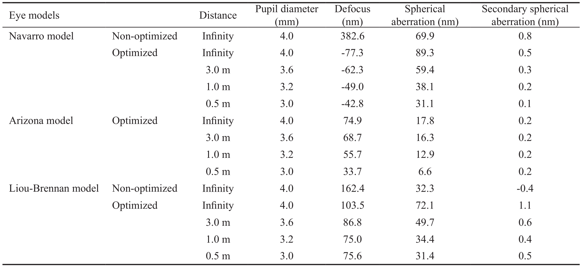
Zernike coefficients of defocus and spherical aberration, both primary and secondary are shown in Table 2. In the Navarro and Liou-Brennan models are obvious the differences before and after optimization at infinity. All models decrease their aberrations as the target approaches, as a result of the pupil diameter decrease and the aberrations that are introduced by the crystalline lens.
The RMS diameter is the root-mean-square error radial size. It is a rough image of the spread of rays on the retinalfield. The Airy disk diameter shows the diameter of the light spot focused on the retina and depends on the diffraction of light through the pupil and its diameter[23-24].
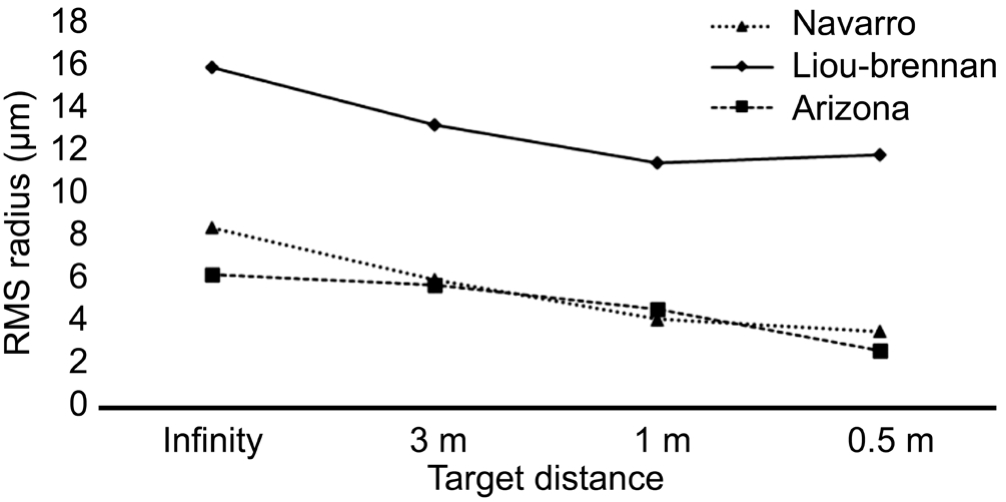
The RMS error and Airy disk diameters of the eye models while the target distance decreases (and the models accommodate)are included in Table 3. The results of Table 3 are graphically presented in Figures 5 and 6.Obviously, the RMS error diameter decrease as the model eye accommodates and the Airy disk diameter increase as the pupil diameter decrease.

Figure 3 Spot diagrams for the three eye models In the Liou-Brennan model the circles of the rays are decentred to the right of the central spotbecause of the decentration of the pupil, creating the characteristic tail of the coma aberration. All dimensions are in μm. In each of the spot diagrams the left one is for 0 angle and the right one for 5 of angle between the incoming light rays and the optical axis.

Figure 4 MTF graphs for the three models The dashed line shows the diffraction limit. OTF: Optical transfer function.
Table 1 Total eye lengths for the optimized Navarro and Liou-Brennan models

The Arizona model does not change its length while accommodating.
DISCUSSION
In the present study there has been a comparison between three schematic eye models (Navarro, Arizona and Liou-Brennan)[2,8-9] in terms of accommodation. The Arizona eye model was able to accommodate by a mathematical algorithm while the other two models had to be changed. These changes included alterations of the radii of curvature of their crystalline lens and the thicknesses of the anterior chamber and the crystalline lens, as the target distance was decreasing. All the parameter changes were selected from the literature. An optimization was implemented in the models with variable the vitreous thickness, in order to get the best image quality in the retina with the chosen parameters in each target distance. A successful accommodation should result in small or no change at all in the vitreous thickness (and the total eye length), while a failure in accommodation should result in unrealistic changes in the total eye length (e.g. larger than 1 mm).
Table 2 The Zernike coefficients for the eye models in all distances of accommodation

Table 3 RMS radius and Airy disk diameter for all eye models in all distances of accommodation

RMS: Root mean square.

Figure 5 RMS error radius over target distance.

Figure 6 Airy disk diameter over target distance.
Letter F Diffraction Images It is obvious from Figure 1 that the optical result on the retina is optimal for all models while accommodating. The Navarro model is a bit myopic, as it is obvious for the far target distances but the image gets clearer as the target distance decrease. The Liou-Brennan model has a characteristic coma blur at the right of the image. The Arizona model shows almost the same retinal image for all distances because it is created to be always optimized through mathematical functions. It doesn’t simulate an average eye but a perfect one. All our results and images are optical simulations of the image as it is refracted on the retina. For the visual results, the neural process that takes place in the brain has to be taken into account, which is not a topic of this paper.
Spot Diagrams and Modulation Transfer Function Graphs In Figures 3 and 4, the comparison shows that the best accommodation is given by the Arizona model. On the other hand,the other two models show quite similar results and in a good accordance to the Arizona. Moreover, the Liou-Brennan model also simulates the characteristic coma “tail” which is a result of the decentration of its pupil.
In Figure 4 the Arizona model shows a better MTF than the other models and is close to be diffraction limited. But it is known that the real eye is far from that.
Total Eye Lengths While accommodating both the Navarro and Liou-Brennan models change their total length while the Arizona model is not. The changes between the accommodation levels are small and comparable between them. Our simulations’results are in the same way with some new studies that have measured the axial length in vivo[18,25], but the difference between our results and theirs is about one magnitude class.This difference is assumed to exist because in our work a merit function was used in order to optimize our models. So there has to be a difference between the merit function of the optical design program and the one that a real human optical system uses.
Aberrations In the aberration results, the Navarro model shows a negative sphere coefficient while the other two models have a positive one. This negative sphere results in a blurry image. All models also have a positive spherical aberration while the secondary spherical aberration is almost zero. It can be observed that the sphere and spherical aberration increase or decrease (in absolute values) in parallel. In other words, the spherical aberration always tries to correct the total sphere that is produced by the cornea and the accommodation process.
According to our results we have observed that the nonaccommodative eye models can simulate accommodation if they are fed with sufficient and correct data. To our knowledge there is no study to compare ourfindings with, but there exists a work in comparing non accommodative eye models[26]. If we compare our findings with this study, then we have to agree that the Liou-Brennan model is more accurate to the biological human eye. It is more detailed by using a gradient refractive index lens and a decentred pupil but this does not make it the perfect model. If this model is selected to simulate a real human eye then more data are needed to be input, in order to be more accurate. There is no model eye that we can propose as the best and the selection depends always on the study,the data that are used and the complexity of the model that is needed.
In the field of the vison science there are many works about accommodating eye models, theoretical and computer designed[10-17] and about how they are created or designed to work.In this study it is shown that, the classical models, even the ones that are not designed to simulate accommodation could be possible tools in research. If they are fed with data that optimize accommodation they can provide simulations which are comparable with the ones of accommodating eye models.So, in a possible input of customized data from a specific subject, they should be able to simulate this customized eye as well. These simulations could be afirst tool in simulating far and near vision for specific applications like spectacle or contact lenses design or fitting. They can also provide some results for blurred vision, halos, light scattering, loss of contrast sensitivity etc. It has to be noted that these can be only thefirst results, and further research, tests and analysis should be considered.
We can conclude that the three models simulated accommodation in good accordance compared between each other.Every difference between the simulations’ results, can be changed by changing the parameters of their optical media.Providing them with more accurate values, we can customize them and get more precise results, maybe without differences between models.
REFERENCES
1 Navarro R. The optical design of the human eye: a critical review. J Optom 2009;2(1):3-18.
2 Schwiegerling J. Field Guide to Visual and Ophthalmic Optics. SPIE Publications; 2004.
3 Rozema JJ, Atchison DA, Tassignon MJ. Statistical eye model for normal eyes. Invest Ophthalmol Vis Sci 2011;52(7):4525-4533.
4 Atchison D, Smith G. Optics of the human eye. Oxford: Elsevier Health Sciences; 2000.
5 Jesus DA, Iskander DR. Simplifying numerical ray tracing for twodimensional non circularly symmetric models of the human eye. Appl Opt 2015;54(34):10123-10127.
6 Polans J, Jaeken B, McNabb RP, Artal P, Izatt JA. Wide-field optical model of the human eye with asymmetrically tilted and decentered lens that reproduces measured ocular aberrations. Optica 2015;2(2):124-134.
7 Artal P, Benito A, Tabernero J. The human eye is an example of robust optical design. J Vis 2006;6(1):1-7.
8 Navarro R, Santamaría J, Bescós J. Accommodation-dependent model of the human eye with aspherics. J Opt Soc Am A 1985;2(8):1273-1281.
9 Liou HL, Brennan NA. Anatomically accurate, finite model eye for optical modeling. J Opt Soc Am AOpt Image Sci Vis 1997;14(8):1684-1695.
10 Navarro R. Adaptive model of the aging emmetropic eye and its changes with accommodation. J Vis 2014;14(13):21.
11 Goldberg DB. Computer-animated model of accommodation and presbyopia. J Cataract Refract Surg 2015;41(2):437-445.
12 Reilly MA. A quantitative geometric mechanics lens model: insights into the mechanisms of accommodation and presbyopia. Vision Res 2014;103:20-31.
13 Van de Sompel D, Kunkel GJ, Hersh PS, Smits AJ. Model of accommodation: contributions of lens geometry and mechanical properties to the
development of presbyopia. J Cataract Refract Surg 2010;36(11):1960-1971.
14 Lanchares E, Navarro R, Calvo B. Hyperelastic modelling of the crystalline lens: Accommodation and presbyopia. J Optom 2012;5(3):110-120.
15 Richdale K, Sinnott LT, Bullimore MA, Wassenaar PA, Schmalbrock P, Kao CY, Patz S, Mutti DO, Glasser A, Zadnik K. Quantification of age-related and per diopter accommodative changes of the lens and ciliary muscle in the emmetropic human eye. Invest Ophthalmol Vis Sci 2013;54(2):1095-1105.
16 Huang JY, Moore D. Computer simulated human eye modeling with GRIN incorporated in the crystalline lens. Journal of Vision 2004;4(11):58.
17 Gambra E, Ortiz S, Perez-Merino P, Gora M, Wojtkowski M, Marcos S. Static and dynamic crystalline lens accommodation evaluated using quantitative 3-D OCT. Biomed Opt Express 2013;4(9):1595-1609.
18 Zhong J, Tao A, Xu Z, Jiang H, Shao Y, Zhang H, Liu C, Wang J.Whole eye axial biometry during accommodation using ultra-long scan depth optical coherence tomography. Am J Ophthalmol 2014;157(5):1064-1069.
19 Kasthurirangan S, Markwell EL, Atchison DA, Pope JM. MRI study of the changes in crystalline lens shape with accommodation and aging in humans. J Vis 2011;11(3).pii:19.
20 Charman WN. The eye in focus: accommodation and presbyopia. Clin Exp Optom 2008;91(3):207-225.
21 Rosales P, Dubbelman M, Marcos S, van der Heijde R. Crystalline lens radii of curvature from Purkinje and Scheimpflug imaging. J Vis 2006;6(10):1057-1067.
22 Dubbelman M, Van der Heijde GL, Weeber HA. Change in shape of the aging human crystalline lens with accommodation. Vision Res 2005;45(1):117-132.
23 Fischer R, Tadic-Galeb B. Optical System Design. Second Edition.New York: The McGraw-Hill Companies, Inc.; 2008.
24 Smith WJ. Modern Optical Engineering: The Design of Optical Systems.Third Edition. New York: The McGraw-Hill Companies, Inc.; 2000.
25 Read SA, Collins MJ, Woodman EC, Cheong S-H. Axial Length Changes During Accommodation in Myopes and Emmetropes. Optom Vis Sci 2010;9(87):656-662.
26 Almeida MSd, Carvalho LA. Different schematic eyes and their accuracy to the in vivo eye: a quantitative comparison study. Braz J Phys 2007;2(37):378-387.