INTRODUCTION
Several studies have evaluated corneal surface changes and postoperative astigmatism after vitreoretinal procedures such as 20-gauge pars plana vitrectomy (20-G PPV)[1-4].Recently, the 25-gauge transconjunctival sutureless vitrectomy(25-G TSV) has been introduced as a more beneficial and less traumatic surgery than the conventional 20-gauge vitrectomy system[5] . The advantages of sutureless small-gauge vitrectomy include technical simplicity without conjunctival dissection and scleral suturing, less traumatic conjunctival and scleral manipulation, reduced operative time, less intraocular in fl ammation and more rapid postoperative visual recovery[6].Several studies showed that the cornea contour is significantly changed, causing postoperative astigmatism, by the 20-gauge standard vitrectomy[7-9]. The induced postoperative astigmatism may be attributed to the cauterization of the sclera and the sutures of the entry sites. In contrast, it has been reported that regular and irregular astigmatism of the cornea did not change in 25-G TSV[10]. To our knowledge, corneal topographic changes and surgically induced astigmatism (SIA) following combined phacoemulsification and 25-G TSV was not investigated before. The importance of our study comes from the fact that, the combined operation is done much more frequently than vitrectomy alone as most of retinal pathologies requiring vitrectomy are present in nearly the same age group of starting senile cataract. Moreover, the diabetic indications of vitrectomy is usually associated with early cataract. In addition, vitrectomy itself causes cataracts to progress. Thus combined phacoemusification and pars plana vitrectomy has been advocated as an effective and safe procedure to handle cases with both cataract and posterior segment diseases[11-12]. In this study, we used videokeratography to analyze topographic changes of the cornea after combined phacoemulsification and 25-G TSV with a follow up period of 6mo to evaluate whether combined self-sealing scleral tunnel incision phacoemulsification (which is also considered to be the least astig-matic[13])and 25-G TSV could sufficiently cause SIA and/or corneal topographic changes to affect postoperative visual outcome.
SUBJECTS AND METHODS
Study Design A retrospective consecutive study was conducted on all patients underwent combined phacoemulsification and 25-G TSV for different vitreoretinal pathologies.
Patients The study was done on 96 eyes of 87 consecutive patients who attended the retinal clinics at Ophthalmology Department, Tokushima University Hospital, Tokushima, Japan from May 2009 to October 2010. The study followed the tenets of Declaration of Helsinki. All patients with a vitreoretinal pathology requiring combined phacoemuslification and 25-G TSV were included with 6mo follow up. Eyes with previous cataract, vitreous surgery or glaucomafiltration surgery were excluded. Those with a histo ry of corneal trauma or corneal disease (dystrophies, de generations, infections, etc.) that might in fl uence corneal topography and eyes which required suturing of the sclerotomy site due to frank leakage were also excluded from the study.
Patients' medical sheets were reviewed for the following parameters: age, gender, best corrected visual acuity (BCVA),anterior segment examination, slit-lamp biomicroscope and corneal topography which is routinely done by the same trained examiner (preoperatively and 2wk, 1, 3, 6mo postoperatively).
Surgical Technique All patients underwent a standardized surgical procedure for 25-G TSV combined with phacoemulsification and intraocular lens (IOL) implantation (acrylic foldable IOL) under retrobulbar anesthesia by 3 surgeons.
Phacoemulsification was done through a superior 3.5 mm self-sealing scleral tunnel incision which was created using a crescent knife and a 3.5 mm keratome at 12 o’clock.The 25-G TSV was done using the newly designed 25-gauge Naito microcannula system (Duckworth & Kent Inc., Hertfordshire, UK)[14]. The conjunctiva was fixed to the sclera with afixation ring and the incisions were made by inserting a 25-gauge microvitreoretinal (MVR) 4 mm posterior to the limbus. The 25-gauge microcannulas were inserted through the scleral tunnel using a cannula inserter.
Core vitrectomy and vitrectomy around sclerotomy sites were done followed by intravitreal injection of triamcinolone acetonide (TA) and then the surgery was completed according to the existing pathology.
After completion of surgery, microcannulas were removed and the conjunctiva was pushed laterally and pressure was applied over the sclera for wound closure for a few seconds. Any leakage would require suture placement.
Corneal Topography Corneal topography was done for all eyes (TMS-2N, TOMEY corporation, Nagoya, Japan) preoperatively and at the 2wk, 1st, 3rd and 6th month postoperatively.Three measurements were taken by a trained examiner at each follow-up visit and the image with the highest quality was analyzed .
Statistical Analysis Visual acuity was recorded in decimal equivalent and then converted to the logarithm of the minimum angle of resolution (logMAR) equivalents for statistical analysis. All parameters were statistically analyzed using the paired t-test; using SPSS software for Windows, with a P value of less than 0.05 was considered to be statistically significant.The topographic parameters analyzed statistically were the average corneal power (ACP), corneal surface cylinder (CSC),surface asymmetry index (SAI) and surface regulatory index(SRI). SIA was calculated by SIA calculator software version 2.1, © 2010, Dr. Saurabh Sawhney, Dr. Ashima Aggarwal.Assuming a SIA of 0.5 diopter would be clinically meaningful.
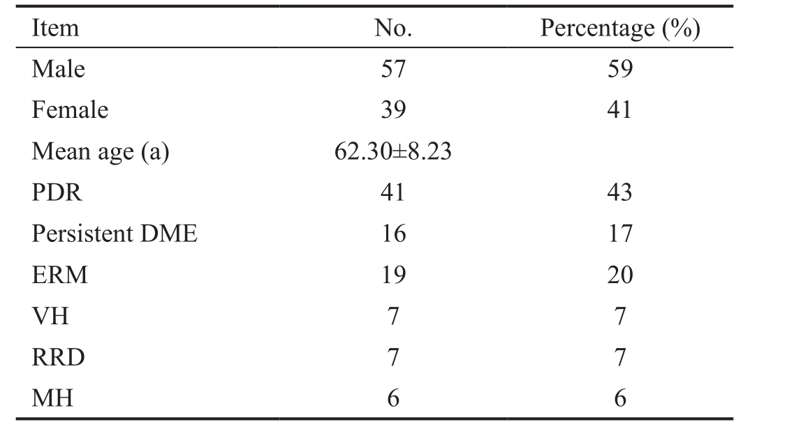
Table 1 Baseline characteristics

PDR; Proliferative diabetic retinopathy; DME; Diabetic macular edema;ERM: Epiretinal membrane; VH: Vitreous hemorrhage; RRD:Rhegmatogenous retinal detachment; MH: Macular hole.
RESULTS
Of the 96 eyes, 57 (59%) were men, 39 (41%) were women,and 47 (49%) were right eyes. Mean age was 62.30±8.23y(range 43-86y). The 41 (43%) had proliferative diabetic retinopathy (PDR), 16 (17%) had persistent diabetic macular edema (DME), 19 (20%) had macular epiretinal membrane,7 (7%) had vitreous hemorrhage, 7 (7%) had rhegmatogenous retinal detachment, and 6 (6%) had idiopathic macular hole(Table 1). Follow-up period was for 6mo.
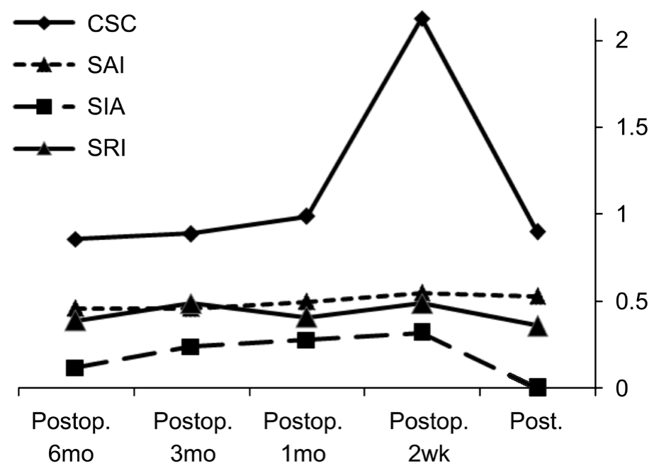
Mean ACP was 43.45±2.44 D preoperatively and 43.77±1.03 D at 2nd week (P=0.168), 43.85±1.02 D atfirst month (P=0.084),43.82±1.02 D at 3rd month (P=0.113), and 43.87±1.05 D at 6th month postoperatively (P=0.655).
Mean CSC was 0.90±0.48 D preoperatively and 2.13±10.53 D at 2nd week (P=0.255), 0.98±0.58 D atfirst month (P=0.826),0.89±0.48 D at 3rd month (P=0.925) and 0.98 ±0.47 D at 6th month postoperatively (P=0.082).
Mean SRI was 0.36±0.35 D preoperatively and 2.13±10.53 D at 2nd week (P=0.006), 0.41±0.30 D atfirst month (P=0.270),0.48±1.22 D at 3rd month (P=0.371) and 0.39±0.29 D at 6th month postoperatively (P=0.70).
Mean SAI was 0.53±0.39 D preoperatively and 0.55±0.28 D at 2nd week (P=0.658), 0.50±0.26 D atfirst month (P=0.498),0.46±0.26 D at 3rd month (P=0.142), and 0.46±0.26 D at 6th month postoperatively (P=0.162) (Table 2).
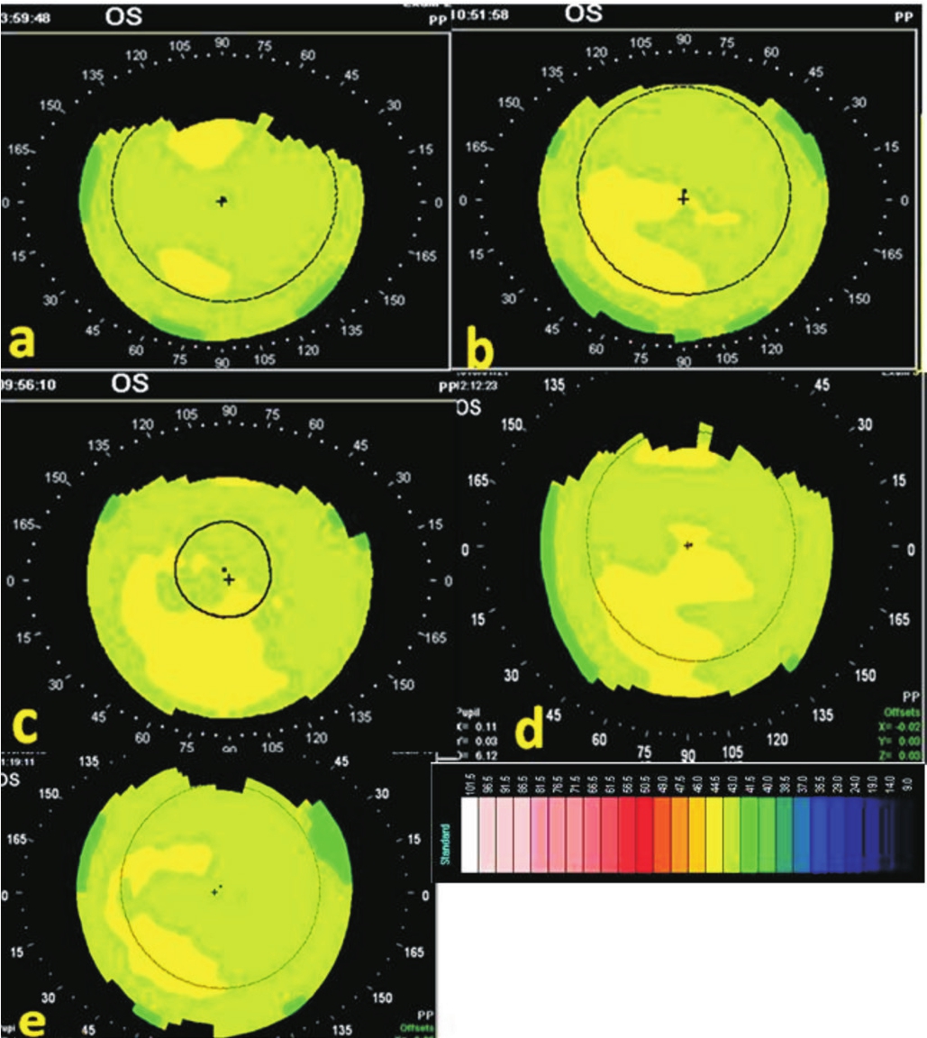
Topographical indices in each follow-up visit are shown in Figure 1. Mean surgically induced astigmatism was 0.3 D at 2wk, 0.28 D at 1mo, 0.24 D at 3rd month and 0.12 D at 6th month postoperatively. Corneal topography of 2 example patients after combined phacoemulsification and 25-G TSV are shown in Figures 2 and 3.The changes in SIA at each postoperative period are shown in Table 2, and Figure 1.
Table 2 Corneal topographic indices in the different follow-up periods

Preop.: Preoperative; Postop.: Postoperative; ACP: Average corneal power; CSC: Corneal surface cylinder; SAI: Surface asymmetry index;SRI: Surface regularity index. aStatistically significant.

Figure 1 Changes in topographic indices in each follow-up visit.

Figure 2 Corneal topography of a patient after combined phacoemulsification and 25-G TSV (Case 1) Preoperative (a), 2nd postoperative week (b), 1st postoperative month (c), 3rd postoperative month (d) and 6th postoperative month (e).
DISCUSSION
Corneal topographic changes and SIA following combined phacoemulsification and vitrectomy can be induced by either procedure[15]. It has always been assumed that scleral incision phacoemulsification would minimize the postoperative astigmatism[13]. Also corneal surface and astigmatic changes are previously reported to be minor in the early postoperative period after 25-G TSV system[10]. To our knowledge, there are no articles which investigate the effect of combined phacoemulsification and 25-G TSV on corneal topography and SIA.

Figure 3 Corneal topography of a patient after combined phacoemulsification and 25-G TSV (Case 2) Preoperative (a), 2nd postoperative week (b), 1st postoperative month (c), 3rd postoperative month (d) and 6th postoperative month (e).
Therefore in our study we evaluated corneal topographic and astigmatic changes 2wk, 1st, 3rd and 6th months following scleral tunnel phacoemul sification combined with 25G-TSV to identify early and late postoperative serial astig matic and topographic changes which had not been investigated before.The greater the SRI and SAI values, the more irregular and asymmetric the corneal curvature[16]. The normal values of SRI and SAI are below 0.70 and 0.54, respectively[17]. Wilson and Klyce[18] have shown that BCVA is significantly correlated with SRI and SAI. The ACP is an area corrected average of the corneal power ahead of the entrance pupil[19-20] and CSC indicates the cylindrical value of the corneal surface.
In our study, no statistically significant difference was noted between preoperative mean ACP, SAI and CSC and either early or late postoperative values. SRI was significantly increased in the 2nd postoperative week only then returned to baseline values thereafter. These results are consistent with those studies done on 23-G[21] and on 25-G TSV (vitrectomy alone) in which no significant changes were observed in SAI,CSC and ACP values within thefirst month postoperatively[5].In our study, the maximum reported SIA is 0.3 D at the 2nd postoperative week which decreased gradually till reach a minimum value at the 6th postoperative month (0.12 D). This is much less than reported previously either following 25-G TSV or 23-G TSV alone in which mean induced astigmatism was reported to be 0.38 D[22] for 25-G TSV and 0.67 D, 0.36 D and 0.33 D for the 1st postoperative day, week and month respectively following 23-G TSV[21]. Our maximum reported SIA also was much less than the minimum reported value of SIA following combined phacoemulsification and 20-G PPV or phacoemulsification and 23-G TSV with 2.2 clear corneal[15].Our results may be attributable to several factors, the first factor may be because our phacoemulsification was done through a scleral tunnel self-sealing 3.2 mm incision which was investigated previously to be nearly astigmatically neutral from the 1st postoperative week[23], also the 25-G TSV system makes a tangential transconjunctival scleral tunnels instead of perpendicular sclerotomies in the 20-gauge systems, lack of scleral suturing and cauterization. Another factor may be the use of the newly designed Naito microcannulas through which the 25-G instruments can be easily interchangeably used through the eye without disturbance in the configuration of the sclerotomy site which allows for more natural and better healing. Thus, it has been suggested that the 25-G TSV combined with self sealing scleral tunnel incision may be one of the best combination of the 2 procedures .
Our study also revealed that, astigmatic changes following this combined operation continued not only for the 3rd postoperative month, which is the maximum follow up period reported in most of the previous studies[13,15,24], but SIA continued to decrease gradually till reach the value of 0.12 D by the 6th postoperative month.
One limitation of this study is the lack of patients who underwent combined scleral tunnel phacoemulsification with both standard 20-G PPV and 23-G TSV as control groups which would lead to more clarification of the differences between these three systems. Nevertheless, the current study is the first which evaluates the corneal topographic changes and SIA in the early and late postoperative periods following combined phacoemulsification and 25-G TSV.
In conclusion, combined 25-G TSV and scleral tunnel phacoemulsification procedure induces minor changes in SAI, CSC,ACP and SRI parameters of the cornea either in the early or late postoperative periods. Surgically induced astigmatism also was the minimum among all previous studies done on the same single or combined procedures making it one of the best combination for patients undergoing cataract and vitrectomy surgery. Minor astigmatic and corneal surface changes are among the most important factors expected to result in rapid visual rehabilitation. Moreover our study revealed that corneal astigmatic changes don't reach its maximum improvement by the 3rd postoperative month only but improvement continues till the 6th postoperative month.
REFERENCES
1 Hayashi H, Hayashi K, Nakao F, Hayashi F. Corneal shape changes after scleral buckling surgery. Ophthalmology 1997;104(5):831-837.
2 Smiddy WE, Loupe DN, Michels RG, Enger C, Glaser BM, deBustros S. Refractive changes after scleral buckling surgery. Arch Ophthalmol 1989;107(10):1469-1471.
3 Goel R, Crewdson J, Chignell AH. Astigmatism following retinal detachment surgery. Br J Ophthalmol 1983;67(5):327-329.
4 Fiore JV Jr, Newton JC. Anterior segment changes following the scleral buckling procedure. Arch Ophthalmol 1970;84(3):284-287.
5 Fujii GY, De Juan E Jr, Humayun MS, Chang TS, Pieramici DJ, Barnes A, Kent D. Initial experience using the transconjunctival sutureless vitrectomy system for vitreoretinal surgery. Ophthalmology 2002;109(10):1814-1820.
6 Lakhanpal RR, Humayun MS, de Juan E Jr, Lim JI, Chong LP, Chang TS, Javaheri M, Fujii GY, Barnes AC, Alexandrou TJ. Outcomes of 140 consecutive cases of 25-gauge transconjunctival surgery for posterior segment disease. Ophthalmology 2005;112(5):817-824.
7 Azar-Arevalo O, Arevalo JF. Corneal topography changes after vitreoretinal surgery. Ophthalmic Surg Lasers 2001;32(2):168-172.
8 Domniz YY, Cahana M, Avni I. Corneal surface changes after pars plana vitrectomy and scleral buckling surgery. J Cataract Refract Surg 2001;27(6):868-872.
9 Slusher MM, Ford JG, Busbee B. Clinically significant corneal astigmatism and pars plana vitrectomy. Ophthalmic Surg Lasers 2002;33(1):5-8.
10 Yanyali A, Celik E, Horozoglu F, Nohutcu AF. Corneal topographic changes after transconjunctival (25-gauge) sutureless vitrectomy. Am J Ophthalmol 2005;140(5):939-941.
11 Chung TY, Chung H, Lee JH. Combined surgery and sequential surgery comprising phacoemulsification, pars plana vitrectomy, and intraocular lens implantation:comparison of clinical outcomes. J Cataract Refract Surg 2002;28(11):2001-2005.
12 Demetriades AM, Gottsch JD, Thomsen R, Azab A, Stark WJ,Campochiaro PA, de Juan E Jr, Haller JA. Combined phacoemulsification,intraocular lens implantation, and vitrectomy for eyes with coexisting cataract and vitreoretinal pathology. Am J Ophthalmol 2003;135(3):291-296.
13 He Y, Zhu S, Chen M, Li D. Comparison of the keratometric corneal astigmatic power after phacoemulsification: clear temporal corneal incision versus superior scleral tunnel incision. J Ophthalmol 2009;210621.
14 Naito T. Transconjunctival sutureless 25-gauge vitrectomy with newly designed microcannulas. J Med Invest 2008;55(1-2):51-53.
15 Park DH, Shin JP, Kim SY. Surgically induced astigmatism in combined phacoemulsification and vitrectomy; 23-gauge transconjunctival sutureless vitrectomy versus 20-gauge standard vitrectomy. Graefes Arch Clin Exp Ophthalmol 2009;247(10):1331-1337.
16 Dingeldein SA, Klyce SD, Wilson SE. Quantitative descriptors of corneal shape derived from computer-assisted analysis of photokeratographs. Refract Corneal Surg 1989;5(6):372-378.
17 Alba-Bueno F, Beltran-Masgoret A, Sanjuan C, Biarnes M, Marin J.Corneal shape changes induced by first and second generation silicone hydrogel contact lenses in daily wear. Cont Lens Anterior Eye 2009;32(2):88-92.
18 Wilson SE, Klyce SD. Quantitative descriptors of corneal topography.A clinical study. Arch Ophthalmol 1991;109(3):349-353.
19 Alimisi S, Miltsakakis D, Klyce S. Corneal topography for intraocular lens power calculations. J Refract Surg 1996;12(2):S309-S311.
20 Maeda N, Klyce SD, Smolek MK, McDonald MB. Disparity between keratometry-style readings and corneal power within the pupil after refractive surgery for myopia. Cornea 1997;16(5):517-524.
21 Yanyali A, Horozoglu F, Macin A, Bozkurt KT, Aykut V, Acar BT,Nohutcu AF. Corneal topographic changes after transconjunctival 23-gauge sutureless vitrectomy. Int Ophthalmol 2011;31(4):277-282.
22 Moon SC, Mohamed T, Fine IH. Comparison of surgically induced astigmatisms after clear corneal incisions of dif ferent sizes. Korean J Ophthalmol 2007;21(1):1-5.
23 Wang J, Chen W, Li J, Zhang D, Sun J, Dong X. A dynamic study on the surgically induced astigmatism after phacoemulsification through three different-sized scleral tunnel incisions. Zhonghua Yan Ke Za Zhi 2000;36(2):91-94.
24 Tseng PC, Woung LC, Tseng GL, Tsai CY, Chou HK, Chen CC, Liou SW. Refractive change after pars plana vitrectomy. Taiwan Journal of Ophthalmology 2012;2(1):18-21.