INTRODUCTION
Viewing and photographing the fundus of the human eye is an important diagnostic method in ophthalmology. It offers the chance for an early diagnosis of diabetic retinopathy,age-related macular degeneration, and many other diseases of the eye. As a consequence, many fundus cameras are not available for developing countries[1]. This is the reason,why it is necessary to use alternative techniques which are easy to use, inexpensive and comfortably transportable to difficultly reachable areas. Such an alternative is the usage
of smartphone in combination with spherical Volk lens with plus twenty diopters (D). The examination of eye fundus in developing countries is in the most of their hospitals absolutely unavailable, mostly due to absence of the specialistophthalmologist.
Within the humanitarian mission project of Department of Ophthalmology, Faculty of Medicine, Comenius University in Bratislava and St. Elizabeth University in Bratislava,Slovakia, we performed ophthalmology screening examination in Mapuordit, South Sudan, from January to February 2015.Patients from this outfield part of South Sudan had before no possibility to be examined by an ophthalmologist, neither was provided basic screening of the eye disorders.
The aim of our study is to present thefirst experience of eye fundus photo documentation by using the plus 20 D spherical Volk lens and a smartphone Lenovo S660, O.S. Android with 4.2 Mpix camera and LED fl ash within the screening project of eye disorders in developing countries.
SUBJECTS AND METHODS
In January and February 2015 within the project in Mapuordit,Lakes state, South Sudan, we performed the basic ophthalmological examination. The equipment consisted of paper form of Snellen chart for illiterate, spectacle frame, system of concave and convex lenses, direct ophthalmoscope, spotlight and magnifier glass. Evaluation of the main functions of vision was made [central visual acuity (CVA), findings at eye subsidiary organs, anterior segment, optical media, the presence of eye fundus reflex and changes at the inner eye surface by direct ophthalmoscopy].
In cases that required documentation offindings was used the alternative option to fundus camera-Smartphone Lenovo in combination with spherical Volk lens (+20 D).
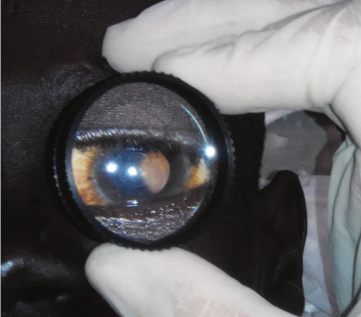
The distance of Volk lens from the investigated eye was about 5 cm and the distance of the lens to smartphone from 25 to 30 cm. The light source was exclusively built-in LED fl ash on smartphone as part of the standard model. Room darkening during the examinations was achieved by the curtains. The images were not adjusted in any way, like rotated, fl ipped or inverted, and were not modified in their sharpness or contrast.The process of photo documentation was performed in artificial mydriasis (phenylephrine plus atropine sulfate Oph. sol.)in laying position in darkened room (Figures 1 and 2). The acquired images were not edited by any additional programs or applications. During the examination patients were not complaining about any discomfort and no complications have occurred.
Table 1 Summary of examined patients including central visual acuity, diagnosis andfindings on the eye fundus

CVA: Central visual acuity; HIV: Human immunodeficiency virus.

Figure 1 Patient and doctor at the beginning of eye fundus examination by smartphone and Volk lens.

Figure 2 Volk lens position by examination of the eye fundus by smartphone.
RESULTS
From the total group of 241 patients (141 males: 58.51% and 100 females: 41.49%) who underwent ophthalmology screening examination performed by one ophthalmologist and one medical student (visual acuity test, anterior segment finding,red eye re fl ex) we recorded ourfindings of the eye fundus by Volk lens and smartphone in patients with transparent optic media. The most common diseases leading to blindness were cataract, trachoma, post-traumatic conditions. Infectious diseases and consequences of untreated infectious diseases were the cause of 20% of the permanent changes on the surface of the eye or the adnexa. In the group of HIV positive patients we did not mention pathologicalfindings on the eye fundus.
Patients, who have had examined eye fundus, where chosen due to their acceptance of examination and transparency of optical media every examination day. In 105 patients (210 eyes) we examined eye fundus by direct ophthalmoscope. In 9 patients we recorded eye fundusfindings by spherical lens and mobile phone. In 96 patients there was no presence of general disease or eye pathology instead of refractive error,therefore they were examined only by ophthalmoscope and documentation of eye fundus was not necessary. Their age was from 16 to 81y, number of female was 47, male 49 and their CVA varied from 0.01 to 1.0.
In the rest group (9 patients) the variety of diagnoses of investigated patients by searching for changes of the eye fundus included Burkitt lymphoma, Kala Azar, malnutrition with unknown etiology, tuberculosis, HIV positive patients,Usher syndrome and hypertension (Table 1). In this group there were 6 males and 3 females, the average age was 32y (from 10 to 62y) and CVA varied from 0.01 to 1.0 (average value 0.8 illiterate chart).
In analyzed group of 9 patients’findings of their eye fundus in 2 patients we succeeded in capturing and we recorded various pigmentations (Figures 3 and 4). In thefirst case we diagnosed a patient with Usher syndrome and we have found small, but numerous pigmentation at the whole eye fundus in the second case, in the patient with hypertension, we recorded changes of vessels typically connected with hypertension. According to ourfirst experience the quality of documentation is sufficient to perform a standard screening of the eye fundus.
DISCUSSION
Disorders of the anterior segment of the eye is possible to capture by digital camera and after, in a short time, contact and discuss the problem with specialist by sending images via email etc[2]. On the other hand, eye disorders that are shown at the back of the inner eye surface were until now impossible to capture. That makes the solution with smartphone and spherical lens very effective and cheap for those, who are in need to make a screening of eye disorders in developing countries[3-4].

Figure 3 Fundus photo performed by smartphone and Volk lens.

Figure 4 Detail of fundus photo of patient No. 7 (Table 1).
There are also other new available technologies allowing more accurate and comfortable screening than fundus cameras. The optics behind fundus photography and the images of the retina that it delivers are relatively straight forward; but actually achieving a high-quality picture can be frustratingly difficult for clinicians. Unfortunately the design of a conventional fundus camera makes it tricky for operators to be sure that the positioning is exactly perfect. The alternative is for the patient to attempt to position the camera themselves and keep it orientated correctly during the imaging procedure. The team at MIT Media Lab has developed a prototype device christened eyeSelfie (as in Eye Self-Imaging), a retinal imaging platform which uses novel optics and an interactive user-interface to provide users with a visual fixation cue indicating when the alignment is correct[5].
Another device able to capture the eye fundus is a smartphone adapter Peek Retina that slips neatly over the in-built camera on your smartphone. The high image quality enables ophthalmologist to view cataracts clearly enough for treatment classification, detect signs of glaucoma, macular degeneration,diabetic retinopathy and signs of nerve disease. Other health problems such as severe high blood pressure and diabetes can also be identified with a good view of the retina[6].
The team of Oluleye performed screening program by preterm infants with birth weight of less than 1.5 kg or gestational age of less than 32wk. In conjunction with the neonatologist,topical tropicamide (0.5%) and phenylephrine (2.5%) was used to dilate the pupils. A pediatric lid speculum was used.Indirect ophthalmoscopy was used to examine the fundus to ensure there were no missed diagnoses. An iPhone 5 with 20 D lens was used to examine the fundus. The App Filmic Pro was launched in the video mode. The camera flash served as the source of illumination. Its intensity was controlled by the App.The 20 D lens was used to capture the image of the retina,which was picked up by the camera system of the mobile phone. They realized by infants, that the images captured by the system were satisfactory for staging and determining the need for treatment in screening for retinopathy of prematurity(ROP) in resource-poor settings[7].
Smartphone-based fundus camera can help clinicians to monitor diseases affecting both central and peripheral retina.It can help patients understand their disease and clinicians convincing their patients regarding need of treatment especially in cases of peripheral lesions. Imaging peripheral retina has not been demonstrated in existing smartphone-based fundus imaging techniques. The device can also be an inexpensive tool for mass screening[8].
Our alternative technique is one of the easiest and requires only basic and accessible equipment-smartphone and Volk lens.With user friendly way of screening we achieved comparable images in a good quality that allowed us to evaluate our results.
CONCLUSION
The examination of eye fundus by using the smartphone and spherical Volk lens with plus 20 D is unpretentious and manageable technique, allowing capture images of the inner eye surface with high quality and reproducibility. These pictures are suitable for screening and photo documentation within ophthalmologic practice. Although in a short time period and difficult conditions during our project in South Sudan, we have examined group of patients with this alternative technique and we realized that this is an alternative way of screening of eye disorders in mission and humanitarian medical ophthalmological projects. Hopefully this technique will become a standard within examination of patients with eye disorders in developing countries.
REFERENCES
1 Michelson G. Teleophthalmology in Preventive Medicine. Berlin:Springer-Verlag Heidelberg; 2015:51.
2 Maamari RN, Keenan JD, Fletcher DA, Margolis TP. A mobile phonebased retinal camera for portable wide field imaging. Br J Ophthalmol 2014;98(4):438-441.
3 Tietjen A, Stanzel BV, Saxena S, Bezatis A, Muller M, Meyer CH. New options for digital photo documentation during routine examination for ophthalmologists. Klin Monbl Augenheilkd 2013;230(6):604-610.
4 Nemcansky J, Kopecky A, Timkovic J, Masek P. The cell phones as devices for the ocular fundus documentation. Cesk Slov Oftalmol 2014;70(6):239-241.
5 Meyer CH. Smart ophthalmologists: smartphones for nothing and the Apps for free? Ophthalmologe 2012;109(1):6-7.
6 Chhablani J, Kaja S, Shah VA. Smartphones in ophthalmology. Indian J Ophthalmol 2012;60(2):127-131.
7 Oluleye TS, Rotimi-Samuel A, Adenekan A. Mobile phones for retinopathy of prematurity screening in Lagos, Nigeria, sub-Saharan Africa. Eur J Ophthalmol 2016;26(1):92-94.
8 Sharma A, Subramaniam SD, Ramachandran KI, Lakshmikanthan C,Krishna S, Sundaramoorthy SK. Smartphone-based fundus camera device(MII Ret Cam) and technique with ability to image peripheral retina. Eur J Ophthalmol 2016;26(2):142-144.