INTRODUCTION
Glaucoma is a leading cause of irreversible blindness worldwide. Thus, early diagnosis of this chronic disease is of crucial importance[1]. Additionally to visual field (VF) tests obtained with standard achromatic perimetry(SAP) as functional parameters, measurements of structural variables at and around the optic nerve head (ONH) have become more and more popular given the significant advances in technology. Especially non-invasively obtained, optical coherence tomography (OCT)-based structural parameters such as the retinal thickness of the posterior pole or the retinal nerve fiber layer (RNFL) thickness can be measured precisely with an accuracy in the range of a few microns. Another huge advantage is the possibility to compare point to point in follow up examinations[2-5].
Recently, the novel OCT-based parameter Bruch’s membrane opening-minimum rim width (BMO-MRW) was introduced.This variable measures the shortest distance from Bruch’s membrane opening (BMO) to the internal limiting membrane[6-8]using radial cross-sectional images through the ONH. The acquisition time for BMO-MRW examinations is longer than for circular RNFL measurements alone and require a better compliance of the examined patients than OCT-based RNFL measurements. BMO-MRW as a structural parameter, together with the established RNFL thickness, has been shown to successfully measure and correlate with the loss of neuroretinal tissue, which happens in glaucoma patients as well as healthy elderly[9]. Yet, the significance or extent of novelty of this relatively new structural parameter BMO-MRW in daily clinical practice remains unclear.
The goal of this prospective study was to evaluate the structural OCT-based parameter BMO-MRW in glaucoma patients and compare the results with RNFL thickness values by correlating both parameters, BMO-MRW and RNFL, to global mean deviation (MD) values of SAP in a clinical routine setting.
SUBJECTS AND METHODS
Patients This prospective cross-sectional study was conducted at the Glaucoma Department of a German University Eye Hospital (Department of Ophthalmology, Technical University of Munich). Inclusion criteria were patients with diagnosed primary open angle glaucoma (POAG) and documented glaucomatous VF defects in SAP who were compared to agematched healthy persons without glaucoma and inconspicuous VF testing (control group). Exclusion criteria for the “glaucoma group” were any non-glaucomatous VF defects, corneal scars,history of any corneal surgery, nystagmus, or any pathology or suspected disease of the ONH or RNFL that may influence VF testing or structural ONH parameters.
All study participants underwent a full ophthalmic examination,including best-corrected visual acuity (BCVA) in decimals obtained with a Snellen projection chart, objective and subjective refraction, slit-lamp biomicroscopy, measurement of intraocular pressure (IOP) with Goldmann applanation tonometry (GAT), gonioscopy, and fundus examination by indirect ophthalmoscopy. The linear vertical cup disk ratio(CDR) was documented. VF testing with SAP was performed prior to dilation of pupils. The study adheres to the tenets of the Declaration of Helsinki (2008) and was approved by the Institutional Review Board.
Standard Achromatic Perimetry VF testing by SAP was obtained for each patient using Octopus 500 G1 (Haag-Streit Diagnostics, Köniz, Switzerland). A reliable VF test was defined as <25% of fixation losses, and <20% false positive and false negative answers. According to the definition of the European Glaucoma Society and previous works by Katz et al[10], a VF defect suspicious for glaucoma was defined as a cluster of at least three or more non-edge-contiguous points with significantly reduced sensitivity (P<0.05), out of these one with a significance of at least P<0.01 on the same side of the horizontal meridian in the pattern deviation plot. All of the included patients in the glaucoma group and none in the control group ful fi lled these criteria.
Optical Coherence Tomography Measurements All included patients were imaged with conventional circular peripapillary ONH cross sectional scans using the Heidelberg Spectralis OCT with an excitation wavelength of 870 nm and 40 000 A-scans per second. Global and six sectorial (superotemporal, temporal, infero-temporal, infero-nasal, nasal, andsupero-nasal) values for both, RNFL thickness and BMOMRW (shortest distance from BMO to the internal limiting membrane) were obtained. The device-specific software-based classification scores for both parameters RNFL and BMOMRW classified each obtained global as well as sectorial value into “within normal limits”, “borderline”, and “outside normal limits”.
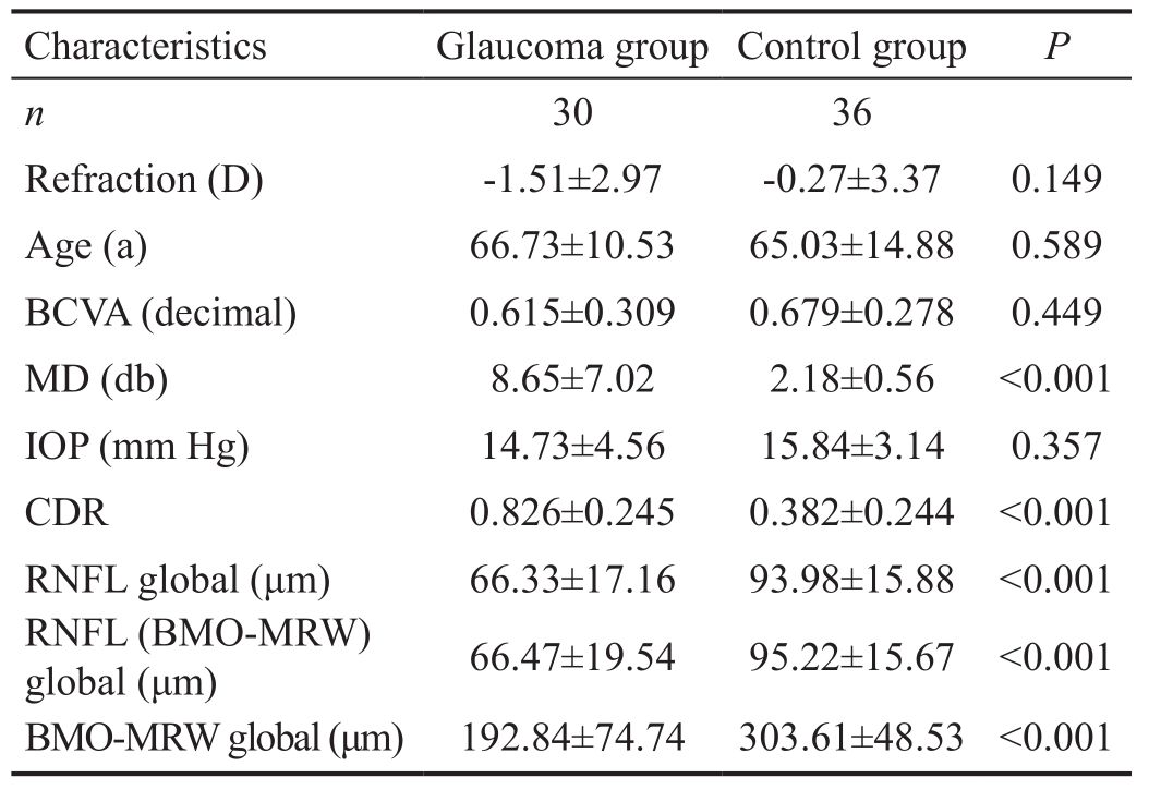
Table 1 Characteristics of glaucoma group and control group

BCVA: Best corrected visual acuity; MD: Mean deviation of standard automated perimetry (SAP) testing; IOP: Intraocular pressure obtained with Goldmann applanation tonometry; CDR: Cup-todisc ratio; RNFL global: Global retinal nerve fiber layer thickness obtained with SD-OCT; RNFL (BMO-MRW) global: Global retinal nerve fiber layer thickness obtained with SD-OCT during BMOMRW measurements; BMO-MRW global: Shortest distance between bruch’s membrane opening and internal limiting membrane.
Structure-function Correlations For further analysis, BCVA and global MD values of the right eye of each patient of the “glaucoma group” and the “control group” were correlated with sectorial and global RNFL and BMO-MRW measurements.
Statistical Analysis Data were collected and analyzed using SPSS Version 22.0 (SPSS Inc., Chicago, IL, USA). The Kolmogorov-Smirnov test was used for testing for normal distribution. Correlation analyses between obtained global functional (SAP) and global as well as sectorial structural (SDOCT) parameters were calculated. Linear regression analyses were performed for comparison between metric and nominal/categorical data.
RESULTS
A consecutive series of 30 glaucomatous right eyes of 30 patients were included in this prospective study and compared to 36 right healthy eyes of 36 individuals in the control group.Patients’ characteristics are given in Table 1. The glaucoma and the control group are age-matched and show similar BCVA and IOP values.
Correlation Analyses We did not find any correlations between BCVA or IOP and the structural OCT-based parameters RNFL or BMO-MRW measurements in the glaucoma group except for the temporal superior quadrant (BCVA and RNFL: Pearson corr. coeff.: 0.399, P=0.029; BCVA and BMO-MRW: Pearson corr. coeff.: 0.447, P=0.013).

Figure 1 Scatter plots between MD (y-axis) and RNFL (x-axis) as well as BMO-MRW (x-axis) values revealing a higher R² for the correlations between MD and BMO-MRW.
Global MD values correlated significantly with global RNFL and global BMO-MRW values in the glaucoma group (Table 2).Correlation analyses between global MD and sectorial RNFL or BMO-MRW values correlated less significantly (Tables 3, 4).Correlation between MD and BMO-MRW was slightly better than between MD and RNFL, as can be seen in Figure 1. In the control group, global MD values did not correlate with global or sectorial RNFL or BMO-MRW measurements.
Subgroup Analysis for Myopia We analyzed the myopic patients (>4 diopters) within the glaucoma group (n=6). Here we found a tendency for better correlation between MD and BMOMRW than between MD and RNFL measurements (Table 5).
DISCUSSION
A clear understanding of the structure-function relationship in glaucomatous eyes remains a challenge and a subject of current and future research. The vast majority of patients with POAG reveal structural changes before functional impairments occur, while in some patients VF changes can either appear simultaneously with RNFL thinning or even precede the observed structural changes[2,11-12]. Despite the attempt to relate function to structure with various models[13-15], it remains of high importance to monitor functional as well as structural parameters in patients with known glaucoma[16].
The recently introduced novel structural parameter BMOMRW measures the shortest distance from BMO to the internal limiting membrane, and is regarded to be a sensitive and stable structural parameter in diagnosis and follow up of glaucoma patients[6,17].
When MD values of VF tests from patients with manifest glaucoma were correlated with both structural OCT-based parameters BMO-MRW and RNFL, we observed that sectorial structure-function analyses were inferior to global correlation analyses. Our observation of a moderate to good structurefunction relationship between global RNFL as well as BMO-MRW and VF sensitivity in the glaucoma group are in agreement with our understanding of glaucoma being a general neurodegenerative disease[18-19].
Table 2 Correlation (Pearson correlation coefficient) between global MD and global OCT-based RNFL and BMO-MRW measurements

aP<0.05 are significant correlations.
Table 3 Correlations (Pearson correlation coefficient) between global MD and sectorial OCT-based RNFL measurements

Temp: Temporal sector; Temp sup: Temporal superior sector; Temp inf:Temporal inferior sector; Nas: Nasal sector; Nas sup: Nasal superior sector; Nas inf: Nasal inferior sector. aP<0.05 are significant correlations.
Table 4 Correlations (Pearson correlation coefficient) between global MD and sectorial OCT based BMO-MRW measurements

Temp: Temporal sector; Temp sup: Temporal superior sector; Temp inf:Temporal inferior sector; Nas: Nasal sector; Nas sup: Nasal superior sector; Nas inf: Nasal inferior sector. aP<0.05 are significant correlations.
Table 5 Correlation (Pearson correlation coefficient) between global MD and global OCT-based RNFL and BMO-MRW measurements of myopic patients (>4 diopters, n=6)

aP<0.05 are significant correlations.
The only exception were the temporal superior and temporal inferior sectors showing a higher structure-function relationship than the other sectors confirming our current understanding that those areas seem to be more sensitive to glaucomatous changes.
When comparing OCT-based RNFL to BMO-MRW, we observed that BMO-MRW showed slightly higher correlations to functional MD values compared to RNFL but interpret those findings only as tendencies and not significant differences.
Our subgroup analysis of myopic patients (>4 diopters, n=6)revealed better correlations between functional VF sensitivity and BMO-MRW compared to RNFL measurements. The temporal superior sector was the only sector to show a constant significant sectorial structure-function relationship, thus again supporting the notion of the temporal sectors of the ONH and peripapillary region to reveal a better structure-function relationship than other sectors in glaucoma patients[20-21].
The varying structure-function correlations between both OCT-based RNFL and BMO-MRW parameters, especially in myopic patients, can be explained with the fact, that for RNFL measurements, the peripapillary circular scan is placed manually around the ONH and does not take into account altered anatomical papillary or peripapillary structures since the prede fi ned diameter and circular shape of the scan remain the same. In contrast, BMO-MRW measurements are based on the actual anatomical opening of Bruch’s membrane of the examined eye and therefore automatically respect structural variations of the evaluated ONHs and their peripapillary structures.
Recent publications have shown a decline of BMO-MRW and RNFL thickness as a natural degenerative process in healthy subjects over time. Interestingly, those changes of BMO-MRW measurements did not highly correlate with the observed changes of RNFL measurements[7]. This observed difference also might point to the above mentioned difference of the two obtained structural OCT-based parameters with BMOMRW being less dependent on structural variations of the ONHs. Future longitudinal studies will have to compare both parameters RNFL and BMO-MRW over time in glaucoma patients with an expected necessity to adjust for age[9] and eventually also for sectors.
A limitation of our one-center study is the relatively small number of patients and its cross-sectional character which does not allow for interpretation regarding possible developments over time. Another limitation is the small number of myopic patients in our subgroup analysis which does not allow to draw strong conclusions independent of the results, but provides a useful hint for a possible better suitability of BMO-MRW compared to RNFL measurements in myopic patients. This careful observation has to be evaluated in larger groups of myopic patients over time.
In summary, we were able to correlate the structural OCT-based parameters RNFL and BMO-MRW to functional VF sensitivity in glaucoma patients. Both parameters RNFL and BMO-MRW seem to be similarly suitable for diagnosis in a clinical setting with BMO-MRW showing higher correlations than RNFL in myopic glaucoma patients.
ACKNOWLEDGEMENTS
Conflicts of Interest: Reznicek L, None; Burzer S, None;Laubichler A, None; Nasseri A, None; Lohmann CP, None;Feucht N, None; Ulbig M, None; Maier M, None.
REFERENCES
1 Quigley HA, Broman AT. The number of people with glaucoma worldwide in 2010 and 2020. Br J Ophthalmol 2006;90(3):262-267.
2 Miglior S, Zeyen T, Pfeiffer N, Cunha-Vaz J, Torri V, Adamsons I;European Glaucoma Prevention Study (EGPS) Group. Results of the European glaucoma prevention study. Ophthalmology 2005;112(3):366-375.
3 Airaksinen PJ, Tuulonen A, Alanko HI. Rate and pattern of neuroretinal rim area decrease in ocular hypertension and glaucoma. Arch Ophthalmol 1992;110(2):206-210.
4 Zangwill LM, Williams J, Berry CC, Knauer S, Weinreb RN. A comparison of optical coherence tomography and retinal nerve fiber layer photography for detection of nerve fiber layer damage in glaucoma.Ophthalmology 2000;107(7):1309-1315.
5 Silverman AL, Hammel N, Khachatryan N, Sharpsten L, Medeiros FA, Girkin CA, Liebmann JM, Weinreb RN, Zangwill LM.Diagnostic accuracy of the spectralis and cirrus reference databases in differentiating between healthy and early glaucoma eyes. Ophthalmology 2016;123(2):408-414.
6 Reis AS, O'Leary N, Yang H, Sharpe GP, Nicolela MT, Burgoyne CF,Chauhan BC. Influence of clinically invisible, but optical coherence tomography detected, optic disc margin anatomy on neuroretinal rim evaluation. Invest Ophthalmol Vis Sci 2012;53(4):1852-1860.
7 Chauhan BC, Danthurebandara VM, Sharpe GP, Demirel S, Girkin CA,Mardin CY, Scheuerle AF, Burgoyne CF. Bruch’s membrane opening minimum rim width and retinal nerve fiber layer thickness in a normal white population: a multicenter study. Ophthalmology 2015;122(9):1786-1794.
8 Gardiner SK, Ren R, Yang H, Fortune B, Burgoyne CF, Demirel S. A method to estimate the amount of neuroretinal rim tissue in glaucoma:comparison with current methods for measuring rim area. Am J Ophthalmol 2014;157(3):540-549.e1-e2.
9 Vianna JR, Danthurebandara VM, Sharpe GP, Hutchison DM, Belliveau AC, Shuba LM, Nicolela MT, Chauhan BC. Importance of normal aging in estimating the rate of glaucomatous neuroretinal rim and retinal nerve fiber layer loss. Ophthalmology 2015;122(12):2392-2398.
10 Katz J, Sommer A, Gaasterland DE, Anderson DR. Comparison of analytic algorithms for detecting glaucomatous visual field loss. Arch Ophthalmol 1991;109(12):1684-1689.
11 Keltner JL, Johnson CA, Anderson DR, Levine RA, Fan J, Cello KE, Quigley HA, Budenz DL, Parrish RK, Kass MA, Gordon MO,Ocular Hypertension Treatment Study Group. The association between glaucomatous visual fields and optic nerve head features in the ocular hypertension treatment study. Ophthalmology 2006;113(9):1603-1612.
12 Hirooka K, Manabe S, Tenkumo K, Nitta E, Sato S, Tsujikawa A. Use of the structure-function relationship in detecting glaucoma progression in early glaucoma. BMC Ophthalmol 2014;14:118.
13 Swanson WH, Horner DG. Assessing assumptions of a combined structure-function index. Ophthalmic Physiol Opt 2015;35(2):186-193.
14 Danthurebandara VM, Sharpe GP, Hutchison DM, Denniss J, Nicolela MT, McKendrick AM, Turpin A, Chauhan BC. Enhanced structurefunction relationship in glaucoma with an anatomically and geometrically accurate neuroretinal rim measurement. Invest Ophthalmol Vis Sci 2014;56(1):98-105.
15 Hu R, Marin-Franch I, Racette L. Prediction accuracy of a novel dynamic structure-function model for glaucoma progression. Invest Ophthalmol Vis Sci 2014;55(12):8086-8094.
16 Malik R, Swanson WH, Garway-Heath DF. Structure-function relationship in glaucoma: past thinking and current concepts. Clin Exp Ophthalmol 2012;40(4):369-380.
17 Hua R, Gangwani R, Guo L, McGhee S, Ma X, Li J, Yao K. Detection of preperimetric glaucoma using Bruch membrane opening, neural canal and posterior pole asymmetry analysis of optical coherence tomography.Sci Rep 2016;6:21743.
18 Vermeer KA, van der Schoot J, Lemij HG, de Boer JF. RPE-normalized RNFL attenuation coefficient maps derived from volumetric OCT imaging for glaucoma assessment. Invest Ophthalmol Vis Sci 2012;53(10):6102-6108.
19 Leaney J, Healey PR, Lee M, Graham SL. Correlation of structural retinal nerve fibre layer parameters and functional measures using Heidelberg Retinal Tomography and Spectralis spectral domain optical coherence tomography at different levels of glaucoma severity. Clin Exp Ophthalmol 2012;40(8):802-812.
20 Reznicek L, Seidensticker F, Mann T, Hubert I, Buerger A, Haritoglou C, Neubauer AS, Kampik A, Hirneiss C, Kernt M. Correlation between peripapillary retinal nerve fiber layer thickness and fundus auto fluorescence in primary open-angle glaucoma. Clin Ophthalmol 2013;7:1883-1888.
21 Reznicek L, Muth D, Vogel M, Hirneiss C. Structure-function relationship between fl icker-defined form perimetry and spectral-domain optical coherence tomography in glaucoma suspects. Curr Eye Res 2017;42(3):418-423.