INTRODUCTION
Diplopia tests are the common methods used to helping with diagnosing ophthalmoplegia. However, single method cannot fulfill the requirement of clinical practice as the variability of diplopia. To quantify diplopia, there are some methods such as Hess weighted score[1] and Hess area ratio[2].
They both were limited in clinical application and research because of their complexity[3-4]. Therefore, we develop a computerized diplopia test, which is based on the principle of Hess screen test and combined the formation and interpretation theory of the red glasses test and Lancaster screen test,with the help of computer technology[5-7]. In our study, we introduce a new interpretation and quantitative method for the computerized diplopia test[8]. The study followed the request of the Declaration of Helsinki and proved by Ethics Committee of the First Affiliated Hospital of Harbin Medical University.
METHODS
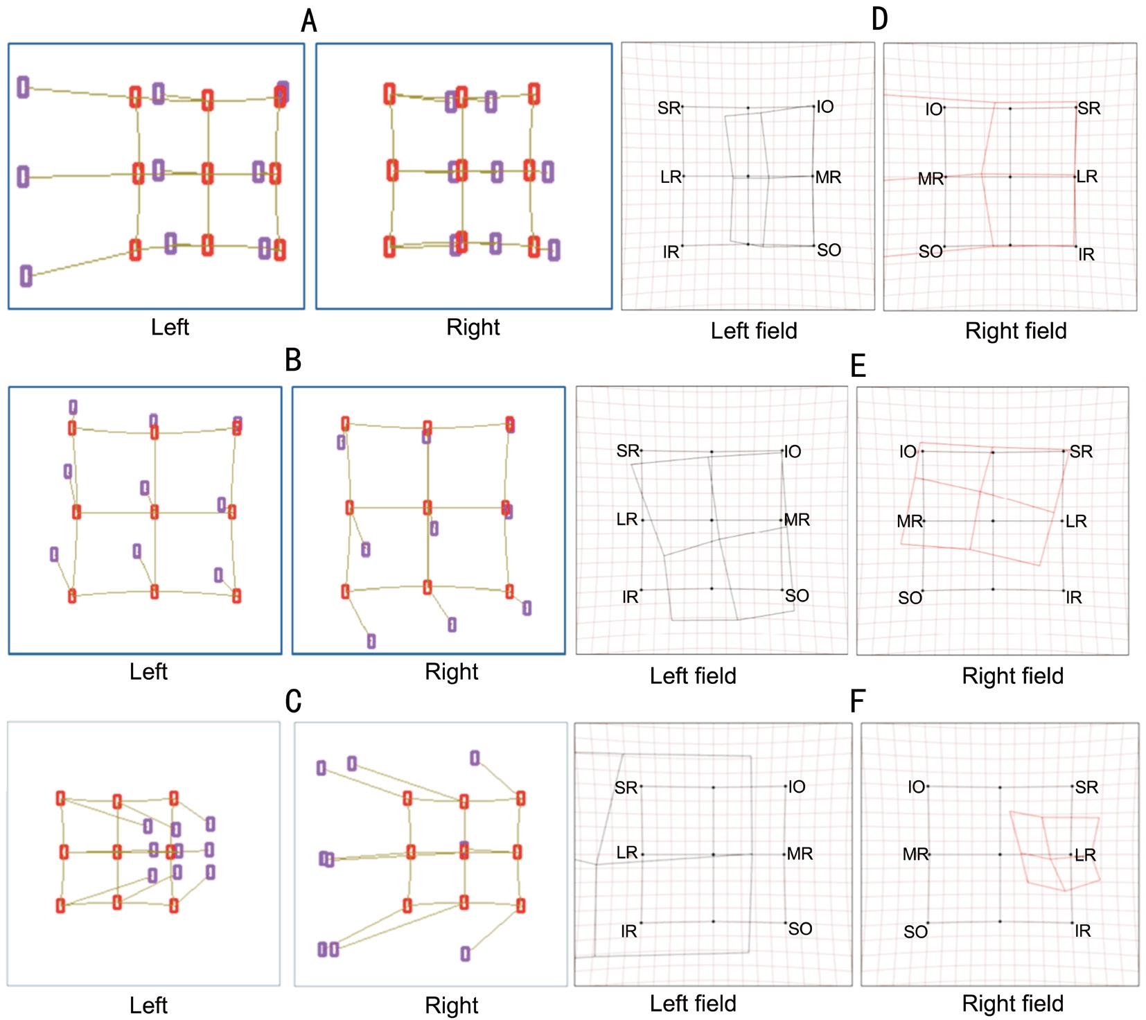
The plot generated by computerized diplopia test reflects the image perceived by patient when the gaze eye is tested. When the affected eye is the gaze eye, the plot is enlarged as the deviated points diffused from standard points. When the health eye is the gaze eye, the plot is lessened as the deviated points shrunken from standard points. There are 9 line segments between the standard point (red point) and perceived point(blue point). They are representing the differences between real image and virtual image in different gaze direction. The directions of these lines show out the direction of diplopia. The length of these lines show out the distance between images.
Interpretation Method We use the diffused plot to determine which eye is affected. The directions of 9 line segments represent the diplopia direction, which indicate the direction of extraocular muscles failed to perform their function and the paralyzed muscle was determined.
Quantitative Method We use the maximum angle of diplopia(MAD) to evaluate severity of diplopia. MAD is the largest deviated angle of the perceived points which is forming the longest line segment in the plot. We use the data from the shrunken plot to avoid inaccuracy caused by the size of screen.
Subjects In our study, we choose patient with ocular motor nerve palsy for clinical verification. The diplopia images of these patients show special pattern. For example, patient with abducent nerve paralysis show the greatest deviation in the outward direction; patient with trochlear nerve paralysis with the dysfunction of superior oblique muscle show great separation in inward and downward direction; patient with oculomotor nerve paralysis with multiple muscles affected show deviations in multiple directions. The plots generate by Hess screen test and the computerized diplopia test show in Figure 1 separately. As we can see, the plots are different from each other. Our new method uses the diffused plot to determine the paralyzed extraocular muscle (Figure 1A-1C). Hess test uses shrunken plot and the location of smallest square area to make a decision (Figure 1D-1F).

Figure 1 Diplopia charts of computerized diplopia test and Hess screen test A-C: Charts generated by computerized diplopia test; D-F:Charts generated by Hess screen test.
We recruited 304 patients with isolated unilateral ocular motor nerve palsy[9] and tested them by computerized diplopia test and Hess screen test. All patients enrolled in our study should with a visual acuity of 20/100 or better and normal retinal correspondence. Patients who received strabismus surgery and patients who incapable of finish the test were excluded. All of the patients have signed consent form. We selected the 20 from the center to eight gaze directions on screen as the standard test mode, with the working distance of one meter. The charts of all patients were randomly divided into 16 groups. In each group,one physician was using two methods, the new interpretation method and Hess interpretation method, to make a diagnosis separately. The physician’s opinions of these two methods were investigated. We use the MAD method and the Hess weighted scoring method[1] to quantify the data of 304 patients then compare consistency of these two methods.
RESULTS AND DISCUSSION

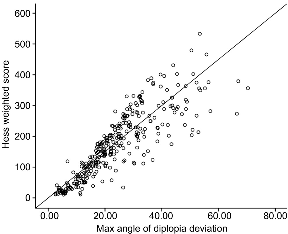
Figure 2 Scatter gram for scores of the two quantitative methods.
Our study enrolled 304 patients with identified diagnosis,91 patients with oculomotor nerve palsy, 35 patients with trochlear nerve palsy and 178 patients with abducent nerve palsy. There are 278 and 257 results consistent with the defined diagnoses with the accuracy rate of 91.44% and 84.53% for the new method and the Hess method respectively. The differences between these methods are statistic significant (P=0.009).Fourteen physicians prefer the new method because it is not only easy to interpret but also intuitive and convenient for clinical use than the Hess method. By Spearman correlation coefficient analysis, the scores calculated by MAD method are positively correlated with the weighted Hess score system(R=0.949, P<0.0001). The results are shown in Figure 2.The computerized diplopia test, as the continue of Lancaster and Lees screen test, is a new method to evaluate diplopia. It is also provide a brand new formation and interpretation method.The direction and length of line segments in the plots generated by computerized diplopia test represented the direction and severity of diplopia. The direction of most serious diplopia represented the direction of extraocular muscles failed to perform their function which used for localization diagnosis.In our study, we compare it with the Hess screen test. The results show that the new interpretation and quantitative method is not only as accurate as the Hess test, but also more suitable for practice. Our study uses the maximum of diplopia from shrunken plot to evaluate severity of diplopia according to the function of yoke muscles. This method could effectively avoid limitation of screen size and it is as accurate as diffuse plot. At the same time, through clinical verification,the results show that MAD is positive correlation with Hess weighted score. According to clinical request and practice and the extraordinary work in developing the computerized diplopia system and interpretation and quantitative methods,we name the new test as Zhou’s Screen test and Zhou’s interpretation and quantitative method. The diagnosis of diplopia is difficult because its complexity and involvement of other clinical departments. We hope this new method which is more convenient and suitable for daily clinical use could assist doctors to make accurate diagnosis and treatment strategy.
ACKNOWLEDGEMENTS
Conflicts of Interest: Zhou LY, None; Liu TJ, None; Li XM,None; Su C, None; Ji XJ, None; Zhao M, None.
REFERENCES
1 Aylward GW, Mccarry B, Kousoulides L, Lee JP, Fells P. A scoring method for Hess charts. Eye (Lond) 1992;6(Pt 6):659-661.
2 Grenga PL, Reale G, Cofone C, Meduri A, Ceruti P, Grenga R. Hess area ratio and diplopia: evaluation of 30 patients undergoing surgical repair for orbital blow-out fracture. Ophthal Plast Reconstr Surg 2009;25(2):123-125.
3 Alhamdani F, Durham J, Greenwood M, Corbett I. Diplopia and ocular motility in orbital blow out fractures 10 year retrospective study. J Cranio Maxill Surg 2015;43(7):1010-1016.
4 Roodhooft JM. Screen tests used to map out ocular deviations. Bull Soc Belge Ophtalmol 2007;(305):57-67.
5 Roper-Hall G. The hess screen test. Am Orthopt J 2006;56(1):166-174.
6 Yoo HS, Park E, Rhiu S, Chang HJ, Kim K, Yoo J, Heo JH, Nam HS.A computerized red glass test for quantifying diplopia. BMC Ophthalmol 2017;17(1):71.
7 Christoff A, Guyton DL. The lancaster red-green test. Am Orthopt J 2006;56(1):157-165.
8 Zhou LY, Liu TJ, Li XM. Adult reference values of the computerized diplopia test. Int J Ophthalmol 2016;9(11):1646-1650.
9 Tamhankar MA, Biousse V, Ying GS, Prasad S, Subramanian PS, Lee MS,Eggenberger E, Moss HE, Pineles S, Bennett J, Osborne B, Volpe NJ, Liu GT, Bruce BB, Newman NJ, Galetta SL, Balcer LJ. Isolated third, fourth, and sixth cranial nerve palsies from presumed microvascular versus other causes:a prospective study. Ophthalmology 2013;120(11):2264-2269.