Dear Editor,
I am Dr. Svetlana V. Jovanovic from the Department of Ophthalmology of the Clinical Centre Kragujevac,affiliated with the Faculty of Medical Sciences, University of Kragujevac, Serbia.
Herein, I present three cases of primary inflammatory choriocapillaropathies (PICCP). PICCP define the spectrum of regrouped diseases where inflammation is on a primary choriocapillaris level, with choriocapillaris nonperfusion and ischemia of the secondary outer retina[1]. The following disease entities are recognized in PICCP, among several others:acute multifocal ischemic choroidopathy (AMIC) in which multiple inflammatory lesions can develop across serpiginous choroiditis (SC), or finally develop into overlapping forms-AMPPiginous choroiditis[2] or has a prolonged progressive clinical course and widespread distribution of lesions described as relentless placoid chorioretinitis (RPC)[1-2]. The etiology is undefined; some suggest an autoimmune/inflammatory cause triggered by an exogenous agent[3-4]. Isolated cases are associated with dengue[5], Epstein-Barr virus (EBV)[2,6],adenovirus (AV)[7], Mycobacterium tuberculosis infection[8],and Lyme borreliosis[9]. The clinical aspect of ocular Lyme borreliosis continues to expand from the initial description of the ocular manifestations of Lyme disease such as severe panophthalmitis, as reported by Steere et al[9] in the 1980s.A number of isolated reports followed, documenting Lyme disease as causing blepharospasm, conjunctivitis, iridocyclitis,optic neuritis, orbital myositis, and strabismus[10]. In this report, we describe three cases of PICCP with expansion and multiplication of the plaques due to Lyme disease.
Ethical Approval Ministry of Health, Republic of Serbia No.500-01-00035, Commission for Health Technology Assessment. The research was performed according to the tenets of the Declaration of Helsinki.
Case 1 One of the possible ocular manifestations of the spirochete Borrelia burgdorferi (B. burgdorferi) was observed in a 47-year-old woman with decreased visual acuity, followed by metamorphopsia in the right eye for a period of one month. While there was no history of a tick bite, physical examination 6mo prior showed typical erythema migrans on the left side of the neck. She was admitted with clinically suspected Lyme disease. On initial examination, the bestcorrected visual acuity (BCVA) was 0.7 (Snellen charts) in the right eye and 1.0 in the left. Intraocular pressure in both eyes was 14 mm Hg. On fundus examination of the right eye in the posterior pole and mid-periphery, we noticed a yellowish-white geographic pattern, confluent in the level of the choriocapillaris and secondary ischemia of the outer retina.Early fluorescein angiography (FA; Visucam FA, Carl Zeiss Meditec AG, Jena, Germany) revealed hypofluoroscent areas indicating choriocapillaris nonperfusion and progressive late staining (Figure 1A, 1B). The clinical presentation indicated the SC form of PICCP[3]. The patient was hospitalized for further diagnostic investigation and treatment. Meanwhile,the positive IgM enzyme-linked immunoassay (ELISA) for B. burgdorferi was confirmed by Western blotting. The results of other laboratory tests (biochemical tests, antinuclear factor,rheumatoid factor, angiotensin-converting enzyme, serum calcium, serum lysozyme levels, lupus anticoagulant, anticardiolipin antibodies and urine calcium tests) were within reference ranges. Tuberculin skin test <5-mm in diameter of induration at 48-72h, and QuantiFERON-TB Gold test was negative. After a few days, on fundus examination of the opposite eye, one yellow-white placoid lesion in the area of both temporal arcades, as well as two changes in the interpapillomacular region, were also observed (Figure 1C).The previously described clinical picture of the opposite eye corresponded to AMIC, but VA was preserved. Antiinflammatory, corticosteroid therapy (60 mg methylprednisolone)under the scheme was added to the doxycycline 2×100 mg over a period of 3wk (Figure 1D). Owing to the progression of bilateral ocular manifestations and a decreased VA in the right eye (BCVA: 0.4), the corticosteroid anti-inflammatory therapy was continued, with gradual tapering, for a period of 3mo (Figure 1C-1E). Three months after the start of therapy,clinical findings were stationary and BCVA was 0.6 in the right eye and 1.0 in the left. We monitored the patient monthly over the first 6mo, and then every 6mo, as there was no disease progression in the opposite eye. After 18mo from disease onset,the funduscopic finding of both eyes remained unchanged.

Figure 1 Bilateral manifestations of the PICCP in a 47-year-old woman A, B: Fundus photo and FA in the SC form where hypofluoroscent areas indicate choriocapillaris nonperfusion and progressive late staining of the right eye; C: Fundus photo of the left eye with AMIC; D, E:After the treatment of oral doxycycline (2×100 mg) daily over a period of 3wk and 60 mg methylprednisolone, tapered gradually, for 3mo.

Figure 2 Multiple areas of fundus photo and FA hyperfluorescency on the mid periphery and posterior pole, capturing the macula of the right eye, only 3d after hospitalization in a 31-year-old man (A, B) and bilateral involvement of the posterior pole (C, D) and the praeequatorial zone (E) after 10d. CFT was 221 μm in the right eye and 175 μm in the left eye (F).
Case 2 A 31-year-old man was hospitalized with the complaint of decreased vision in his right eye for a few days.The patient denied any previous ophthalmic diseases and febrile or flu-like episodes. On initial examination, VA was 0.05 (Snellen charts) in the right eye and 1.0 in the left. Color vision (Ishihara test) in the left eye was preserved. Intraocular pressure in both eyes was 16 mm Hg. The anterior segments were without exudation, and there were no vitreous cells.Fundus examination showed multiple creamy-white lesions at the level of the choriocapillaris, with an inflamed retina above,on the mid periphery, posterior pole, and praeequatorialis.Additionally, in the right eye, the macula was damaged by the inflammation. Given the suspicion of PICCP, we continued with additional tests after the hospitalization. The perimetry figure showed an absolute absence of sensitivity in the right eye, and objective, multifocal scotoma decreased sensitivity in the left eye. An FA showed an area of subretinal placoid lesions in the early stages by visible hypofluoroscency, which indicated choriocapillaris nonperfusion; in the late stages of the disease, hyperfluorescence was seen depending on the severity of the ischemic process. The previously described changes were observed in both eyes in the region of the equatorial zone. In the right eye, the changes had much more extensive involvement of the macula (Figure 2A-2E).Heidelberg retinal tomography (HRT) did not detect structural changes in either eye. Visual evoked potentials (VEP) showed appropriate cortical response in the left eye, while no response was generated in the right eye. Optical coherence tomography(Cirrus OCT, Carl Zeiss Meditec AG, Jena, Germany) of the right eye showed macular atrophy with central field thickness(CFT) of 221 μm. CFT of the left eye was 175 μm (Figure 2).Biochemical and serological test results were within reference ranges. IgM ELISA and Western blot revealed the presence of concomitant acute Borreliosis infection. After the previous examination, the patient was diagnosed with AMIC in the left eye and lesions between the AMIC and SC with relentless evolution in the right; B. burgdorferi was the etiological factor.Systemic antibiotic therapy (doxycycline, 2×100 mg) for 3wk with corticosteroid therapy (prednisolone, 1 mg/kg) under the scheme was prescribed for a period of 3mo. After the antibiotic therapy, the BCVA in the left eye was 1.0, but there was no improvement in the VA of the right eye because of the damage to the retinal pigment epithelium (RPE) of the macular region,recorded by OCT examination (Figure 2).

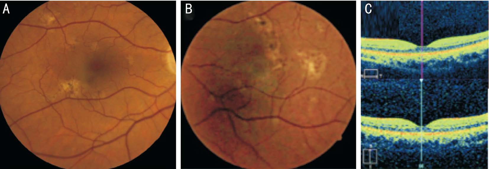
Figure 3 Fundus photo of the right (A) and left eye (B) showed numerous scars typical of placoid lesions after AMIC in a 63-year-old woman and OCT of the same patient. The central macula appears unaffected on OCT (C).
Case 3 A 63-year-old woman came for an ophthalmic examination because of a scotoma appearance in the visual field, about 6mo previously. The patient denied any previous ophthalmic diseases, but revealed that she was bitten by a tick 10mo ago. The patient showed positive results for B. burgdorferi on ELISA and Western blotting. She was treated with doxycycline (2×100 mg) for over a month,by an infectious disease specialist. After completing the treatment for Lyme disease, she noticed her peripheral vision decreasing. On initial examination, the BCVA was 1.0 in both eyes. The anterior segment and vitreous cavity were without inflammatory signs. Fundus examination revealed numerous scars typical of placoid subretinal choriocapillaris lesions after AMIC in both eyes (Figure 3A, 3B). These lesions involved the mid, far peripheral, and posterior pole. The macular area was preserved on OCT examination (Figure 3C).The perimetry showed multifocal scotomas with decreased sensitivity in both eyes, corresponding to our funduscopic findings and the patient’s symptoms. Biochemical testing,serological and immunological identification of infectious agents, and autoimmune status were complemented by multimodal imaging and functional parameters. In this case,B. burgdorferi had already damaged the choriocapillaris, but VA was preserved, so no further treatment, other than patient monitoring, was necessary.
DISCUSSION
Three cases presented three clinical courses of the PICCP in different stages of the disease. Clinical course and visual prognosis depend on initial localization and distribution of nonperfused area in the choriocapillaris. All three patients are characterized by a viral prodrome. Our first patient presented with deep discoloured lesions as AMIC, leading to a progressive course with the form of SC. Progressive,irreversible RPC and reduced immunity was described in the second case report. Earlier changes from AMIC syndrome,consequently after the Borrelia infection, were detected in the third case report.
The etiopathogenesis of the PICCPs remains unclear. Several theories have been described in literature. In 1968, Gass[11]established that placoid lesions at the level of the pigment epithelium and choroid, their development and resolution represented an acute pigment epithelial response to local infectious/noxious agents. Later, Van Buskirk et al[12] suggested choriocapillaris perfusion as the underlying disorder. Other data stated choriocapillary hypoperfusion with secondary changes of the RPE as the primary cause[13]. The cause of this hypoperfusion may be vascular changes of immune origin,which would correlate with human leukocyte antigen (HLA)-DR2, HLA-B7, HLA-A3, and HLA-C7 positivity[14]. In addition, B. Burgdorferi can generate an immune response that is expressed as choriocapillaropathies[15]. A clinical patern described as AMIC-like picture and serpiginous-like choroiditis was seen in a patient with choroidal tuberculosis and Lyme disease[15-19].
Diagnosis and treatment of ocular manifestations seen in the early and late stages of Lyme disease can be facilitated by biochemical and serological examinations complemented by functional parameters and multimodal imaging[3]. Multimodal imaging includes FA, indocyanine green angiography (ICGA)[20], fundus autofluorescence (FAF), OCT[21], and HRT[22].
In our cases, FA provides information regarding the localization and extent of posterior segment inflammatory diseases[23].OCT angiography could help us in the differential diagnosis of PICCP, suggesting that inner choroidal ischemia is the etiology of PICCP[23]. Diagnostic laboratory workup included erythrocyte sedimentation rate, complete blood cell count,and biochemistry. Serological tests for syphilis, human immunodeficiency virus, varicella zoster virus, herpes simplex virus types 1 and 2, Mycoplasma pneumoniae, Mycobacterium tuberculosis, cytomegalovirus, adenovirus, Epstein-Barr virus, toxoplasmosis, mumps, and rubella were also within reference ranges. Presence of B. burgdorferi was confirmed by positive IgM ELISA and Western immunoblotting. Other laboratory tests were within normal ranges. There was no history of previous exposure to tuberculosis and no systemic signs or symptoms that were suggestive of sarcoidosis.Chest radiography showed no indication for sarcoidosis and tuberculosis. The treatment should be adjusted to the course of the inflammatory disease and its etiology.
The treatment of a patient with a known etiology of Lyme disease as the cause of PICCP should be adapted to the course of inflammatory diseases[24]. Patients should be hospitalized,followed by laboratory-proven borreliosis, and introduction of oral antibiotics for 2-3wk: doxycycline 100 mg twice a day, or 500 mg of amoxicillin three times a day[24] or ceftriaxon 2 g/d intravenous[15]. In children, pregnant women, or people with hypersensitivity to tetracycline or erythromycin, penicillin is administered at a dose of 500 mg, 4 times a day[24]. The majority of authors do not recommend therapy for AMIC with a noninfectious etiology[14,24]. The patients were prescribed corticosteroids (topical and general) in infectious PICCP cases with Lyme disease and loss of function[25]. In two of our cases, with the capture of the central region and low VA of the eye, general corticosteroid therapy was recommended,with good therapeutic effect. Initially, the first patient was not treated with corticosteroids because clinical patern resulted in macular atrophy. When the second eye of the same patient was involved, 1 mg/kg/d dose of corticosteroids allowed FA-monitored healing of the satellite lesions. Therefore, if the disease is progressing and function lost, corticosteroid therapy should be initiated. In case 2, with applied intensive corticosteroid anti-inflammatory therapy, a regression of the inflammatory reaction occurred to full scope, with improvement of visual acuity in the better eye. The complete deterioration of visual acuity in the more afflicted eye corresponds to permanent changes of the RPE in the macula.In case 3, we did not recommend therapy, because the patient was already being treated with antibiotics by an infectious disease specialist.
CONCLUSION
PICCP are caused by an inflammation producing choriocapillaris nonperfusion and have characteristic local retinal and subretinal lesions on fundus examination. Differences on fundus examination determine the form of PICCP. In our cases, the clinical course and serological analysis suggested that B.burgdorferi was a possible etiological factor. B. burgdorferi can generate an immune response that is expressed as choriocapillaropathies. Commonly used antibiotics in the treatment did halter the progression of the disease. Adding corticosteroids showed effectiveness in some of our patients.However, even with added anti-inflammatory therapy, the prognosis was not promising if the inflammatory process had caused permanent damage to the RPE in the macula.The analysis of the above-mentioned cases regarding the localization of lesions and foudroyant course of current disease in B. burgdorferi seropositive patients after a tick bite indicates that detailed funduscopic examination is mandatory, as well as timely implementation of therapies.
ACKNOWLEDGEMENTS
Jovanovic SV designed the study, wrote the manuscript.Petrovic NT taught all the procedure of the study. Sarenac Vulovic TS designed the study. Toncic ZG designed the study.Conflicts of Interest: Jovanovic SV, None; Petrovic NT,None; Zivkovic MLJ, None; Toncic ZG, None; Sarenac Vulovic TS, None.
REFERENCES
1 Herbort CP. Inflammatory choriocapillaropathies: rare, intermediary and unclassifiable forms. In: Gupta A, Gupta V, Herbort CP, Khairallah M(eds). Uveitis, Text and Imaging. New Delchi:Jaypee; 2008:90-99.
2 Jones BE, Jampol LM, Yannuzzi LA, Tittl M, Johnson MW, Han DP,Davis JL, Williams DF. Relentless placoid chorioretinitis a new entity or an unusual variant of serpiginous chorioretinitis? Arch Ophthalmol 2000;118(7):931-938.
3 Cimino L, Mantovani A, Herbort CP. Primary inflammatory choriocapillaropathies. Pleyer U, Mondino B, editors. Uveitis and Immunological Disorders. Germany:Springer; 2007:206-228.
4 Crawford CM, Igboeli O. A review of the inflammatory chorioretinopathies:the white dot syndromes. ISRN Inflamm 2013;2013:783190.
5 Goldhardt R, Patel H, Davis JL. Acute posterior multifocal placoid pigment epitheliopathy following dengue fever: a new association for an old disease. Ocul Immunol Inflamm 2016;24(6):610-614.
6 Janani MK, Malathi J, Biswas J, Sridharan S, Madhavan HN. Genotypic detection of epstein barr virus from clinically suspected viral retinitis patients in a Tertiary Eye Care Centre, India. Ocul Immunol Inflamm 2015;23(5):384-391.
7 Thompson SP, Roxburgh ST. Acute posterior multifocal placoid pigment epitheliopathy associated with adenovirus infection. Eye (Lond)2003;17(4):542-544.
8 De Luigi G, Mantovani A, Papadia M, Herbort CP. Tuberculosisrelated choriocapillaritis (multifocal-serpiginous choroiditis): follow-up and precise monitoring of therapy by indocyanine green angiography. Int Ophthalmol 2012;32(1):55-60.
9 Steere AC, Duray PH, Kauffmann DJ, Wormser GP. Unilateral blindness caused by infection with the Lyme disease spirochete, Borrelia burgdorferi. Ann Intern Med 1985;103(3):382-384.
10 Howlett JM, Booth AP. Ocular inflammation as a manifestation of Lyme borreliosis. BMJ 2012;345:e4721.
11 Gass JD. Acute posterior multifocal placoid pigment epitheliopathy.Arch Ophthalmol 1968;80(2):177-185.
12 Van Buskirk EM, Lessell S, Friedman E. Pigmentary epitheliopathy and erythema nodosum. Arch Ophthalmol 1971;85(3):369-372.
13 Rachel R. Caspi RR, Dick A, et al. Immunology of uveitis.Germany:Springer; 2016:39-81.
14 Baxter KR, Opremcak EM. Panretinal acute multifocal placoid pigment epitheliopathy: a novel posterior uveitis syndrome with HLA-A3 and HLA-C7 association. J Ophthalmic Inflamm Infect 2013;3(1):29.
15 Wilkos-Kuc A, Biziorek B, Zarnowski T. Acute posterior multifocal placoid pigment epitheliopathy (APMPPE)--a report of three cases. Klin Oczna 2012;114(4):286-291.
16 Eandi CM, Neri P, Adelman RA, Yannuzzi LA, Cunningham ET Jr;International Syphilis Study Group. Acute syphilitic posterior placoid chorioretinitis: report of a case series and comprehensive review of the literature. Retina 2012;32(9):1915-1941.
17 Bansal R, Gupta A, Gupta V, Dogra MR, Sharma A, Bambery P. Tubercular serpiginous-like choroiditis presenting as multifocal serpiginoid choroiditis. Ophthalmology 2012;119(11):2334-2342.
18 Al Mousa M, Koch F. Acute Borrelia infection inducing an APMPPE-like picture. J Ophthalmic Inflamm Infect 2016;6(1):22.
19 Bansal R. Sharma A, Gupta A. Intraocular tuberculosis. Expert Rev Ophthalmol 2012;7(4):341-349.
20 Agrawal RV, Biswas J, Gunasekaran D. Indocyanine green angiography in posterior uveitis. Indian J Ophthalmol 2013;61(4):148-159.
21 Dolz-Marco R, Rodrigez-Ratón A, Hernández-Martinez P, Pascual-Camps I, Andreu-Fenoll M, Gallego-Pinazo R. Macular retinal and choroidal thickness in unilateral relentless placoid chorioretinitis analyzed by swept-source optical coherence tomography. J Ophthalmic Inflamm Infect 2014;10(2):4:24.
22 Mantovani A, Giani A, Herbort CP Jr, Staurenghi GG. Interpretation of fundus autofluorescence changes in choriocapillaritis: a multi-modality imaging study. Greafes Arch Clin Exp Ophthalmol 2016;254(8)1473-1479.
23 Kuznetcova T, Jeannin B, Herbort CP. A case of overlapping choriocapillaritis syndromes: multimodal imaging appraisal. J Ophthalmic 2012;7(1):67-75.
24 Herbot CP. Primary inflammatory choriocapillarpathiies. Intraocular inflammation. Germany:Springer; 2016:209-228.
25 Kubicka-Trzaska A, Oleksy P, Karska-Basta I, Romanowska-Dixon B.Acute posterior multifocal placoid pigment epitheliopathy (APMPPE)-a therapeutic dilemma. Klin Oczna 2010;112(4-6)127-130.