INTRODUCTION
With the development of corneal refractive surgery and the introduction of wavefront-guided ablation and aspheric ablation, visual quality has become an increasingly important criterion. However, correct alignment of the corneal ablation is crucial to achieving good visual results because decentered optical zones can lead to a signif i cant increase in higher-order aberrations[1], with a decrease in quality of vision,diplopia[2], decreased contrast sensitivity, and night vision disturbances[3]. There are three common choices for corneal ablation centration: centration of the pupil [line of sight(LOS)], the geometric center of the cornea (cornea vertex,CV), and the visual axis of the cornea ref l ex points [coaxially sighted corneal light reflex (CSCLR), also referred to as the“visual axis” in some studies][4-5]. The LOS is defined as the line joining the fi xation point with the center of the entrance pupil[6]. The visual axis is defined as the line joining the fi xation point and the fovea[6].
Early refractive outcomes of laser in situ keratomileusis(LASIK) centered on the CSCLR or the LOS showed that both strategies were safe and effective[7-8]. However, there is still some controversy as to where it is best to center the corneal ablation: on the entrance pupil center (LOS) or on the corneal vertex (an approximate proxy for the visual axis).The LOS is based on the theory that only the bundle of rays of light delimited by the entrance pupil enters the eye, and the LOS represents the chief ray of that bundle of light reaching the fovea[9]; thus, the intersection of the LOS with the cornea should be the desired centration point. In contrast, CSCLR supporters propose that the best optical results are achieved by centering corneal ablation on the corneal vertex, which best approximates the corneal intercept of the visual axis[10].
Among the three ablation centers, the CSCLR distance to the visual axis is approximately 0.02 mm, whereas the other two ablation centers are relatively larger. Hence, practically,it is diff i cult to locate the optic axis at the intersection of the corneal surface, so that the CSCLR is the closest to the “ideal”anatomical point[11-12]. Theoretical modeling indicates that decentration of 0.10 mm can induce aberrations rather than reduce aberrations during myopic wavefront-guided treatments.Centration on the LOS is not ideal strategy, because it defaults the shift in pupil center with differing light conditions[13]. In the present study, we compared the postoperative outcomes with myopic LASIK centered on the LOS or the CSCLR. We evaluated the eff i cacy and safety indexes, refractive outcomes,postoperative wavefront aberrations, contrast sensitivity, and objective visual quality in eyes after femtosecond laser surgery.
SUBJECTS AND METHODS
General Information This randomized, double-masked study compared visual, refractive, and the corneal high-order aberrations, and contrast sensitivity outcomes of the LOS versus the CSCLR with myopic LASIK. In total, 481 eyes of 241 patients (131 males, 110 females), ranging in age from 18 to 35 years old, underwent LASIK surgery between March and September 2014 at Ruijin Hospital, affiliated with Shanghai Jiaotong University School of Medicine. Patients were divided into two groups depending on the different ablation centers:122 patients (243 eyes) underwent treatment with centration on the CSCLR (CSCLR group) and 119 patients (238 eyes)underwent treatment with centration on the LOS (LOS group).The two ablation methods were randomized using a randomnumber table at the inclusion visit.
Inclusion criteria were as follows: age 18-35 years old,preoperative spherical refraction of -2.00 to -12.00 D, refractive cylinder of less than -3.00 D, a stable refractive state for 2y,an intraocular pressure (IOP) of <21 mm Hg, and at least 4wk or 2wk without hard or soft contact lenses, respectively.Exclusion criteria included the following: a history of systemic autoimmune disease, a history of diabetes, other ophthalmic disorders, a history of ocular trauma, and a surgical history.Prior approval by ethic committee of Ruijin Hospital (Shanghai Jiaotong University School of Medicine) was obtained according to the Declaration of Helsinki. All patients were informed of the surgical procedures and use of equipment, and all provided written informed consent.
Methods Patients underwent a full eye examination before LASIK surgery, including evaluations of their uncorrected visual acuity (UCVA), best-corrected visual acuity (BSCVA),IOP, corneal curvature, corneal diameter, corneal thickness,anterior and posterior corneal surface height, corneal topography, refraction and slit-lamp anterior segment, fundus examinations, contrast sensitivity measurement (CSV-1000,VectorVision), and corneal high-order aberrations measurement(Pentacam, Oculus, Germany).
Patients were divided into two groups based on the center of ablation. The surgery procedure was as follows. Topical anesthesia and 2% propoxyphene tetracaine eye drops were applied, along with routine disinfection and shop towels.An eyelid holder was used to open the eye. A suction ring was applied to the eye to hold it in place. Surgery using the femtosecond laser was used to create the corneal fl ap. Adjusted ablation was offset with the ESIRIS excimer laser software.
The femtosecond laser had a bed energy of 0.65 μJ, with a side-cut energy of 0.8 μJ and a repetition frequency of 200 kHz.Myopia stromal ablations were performed using an Allegretto Wave Eye-Q excimer laser (Wavelight Company, Germany).The wavelength and energy intensity of the excimer laser were 193 nm and 180 mJ/cm2, respectively. The corneal ablation was centered on CSCLR or LOS. For the CSCLR group,during surgery the patient subject-fixated coaxially sighted corneal light reflex, the surgeon used left eye to view right corneal light ref l ex and removed the ablation center (red point)to fixation point (green point), whereas the opposite. The vector of pupil shift were calculated from the horizontal and vertical magnitude (X and Y value) according to the following formula.
Vector of pupil shift![]() For example, the ablation center was shifted nasoinferiorly on the x axis by 0.31 mm and 0.12 mm on the y axis, corresponding to an offset of 0.33 mm. For the LOS group, the centering procedure was entirely automated selecting the pupil center and did not rely on surgeon adjustment.
For example, the ablation center was shifted nasoinferiorly on the x axis by 0.31 mm and 0.12 mm on the y axis, corresponding to an offset of 0.33 mm. For the LOS group, the centering procedure was entirely automated selecting the pupil center and did not rely on surgeon adjustment.
At 1, 3, and 6mo postoperatively, follow-up measurements included auto refractometry, manifest refraction, BSCVA,UCVA, Pentacam topography, and corneal high-order aberrations, as well as contrast sensitivity.
Statistical Analysis Data analyses were performed using SAS software (version8.2, SAS Institute, Inc.). Normality of all data samples was fi rst checked using the Kolmogorov-Smirnov test. When parametric analysis was possible, the Student’s t-test for paired data was performed for all parameter comparisons. When parametric analysis was not possible, the Wilcoxon rank-sum test was applied to assess the signi fi cant of differences. For all statistical tests, a Ρ value less than 0.05 was considered statistically signi fi cant.
Table 1 Preoperative parameters
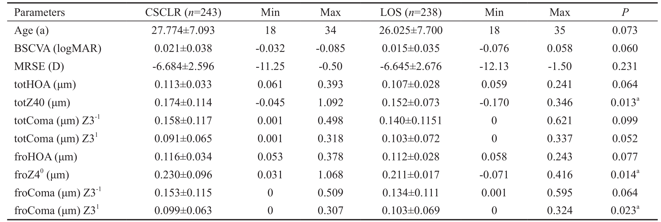
CSCLR: Coaxially sighted corneal light ref l ex; LOS: Line of sight; UCVA: Uncorrected visual acuity; BSCVA: Best spectacle-corrected visual acuity; MRSE: Manifest refraction spherical equivalent; HOAs: Higher-order aberrations, whole corneal spherical aberration (totHOA),corneal anterior surface aberration (froHOA). aStatistically signif i cant difference using the Wilcoxon rank-sum test with continuity correction,Ρ<0.05.
Table 2 Contrast sensitivity data (cd/m2) preoperatively in the two groups in different environments (100%, 25%, 10%, and 5%)

CSCLR: Coaxially sighted corneal light ref l ex; LOS: Line of sight. Statistically signif i cant (Ρ<0.05).
RESULTS
The preoperative parameters for both groups are shown in the Table 1. Preoperatively, the mean age was 27.77±7.1y in the CSCLR group and 26.03±7.70y in the LOS group;Preoperatively the MRSE was -6.68±2.60 D in the CSCLR group and -6.65±2.68 D in the LOS group.
The two groups showed differences in preoperative corneal anterior surface and corneal spherical aberration with MRSE(-0.25 to -0.29 D, -0.30 to -0.59 D, -0.60 to -0.89 D, -0.90 to-12.00 D).
Table 2 shows the preoperative contrast sensitivity function was no statisticaly signif i cant difference in the two groups.
Regarding the distance between the pupil center point and the visual axis preoperation in the two groups, in the CSCLR group, the preoperative distance between the pupil center and the visual axis was 0.1867±0.0925 (0.0141-0.5701) mm,whereas in the LOS group, it was 0.2029±0.109 (0.01-0.5235)mm. There was no signif i cant difference between the groups(Ρ=0.0801; Figure 1).
Postoperative Variables
Postoperative uncorrected visual acuity At 1mo postoperatively, UCVA was 0.009±0.007 in the CSCLR group and 0.001±0.007 in the LOS group (Ρ=0.22). There was no signif i cant difference at 3 or 6mo postoperatively (Figure 2).
Postoperative best spectacle-corrected visual acuity At 1mo postoperatively, BSCVA was -0.007±0.045 in the CSCLR group and -0.012±0.045 in the LOS group (Ρ=0.23). Moreover,117 eyes showed no change in either group. However, in the CSCLR group, 119 eyes gained one line and six eyes lost one line, and one eye lost two lines. In the LOS group, 104 eyes gained one line and 14 eyes lost one line, and six eyes lost two lines. There was no change at 1, 3, or 6mo postoperatively(Figure 3).
Postoperative manifest refraction spherical equivalent Regarding postoperative MRSE, at 1mo postoperatively,the MRSE values of 232 eyes (95%) were within ±0.5 D in the CSCLR group, and 224 eyes (94%) in the LOS group,respectively. There was no signif i cant difference at 3 or 6mo postoperatively (Figure 4).
Table 3 shows the postoperative parameters for both groups.No statistically significant differences were noted between groups in the UCVA, BSCVA, SI, EI and MRSE.
Regarding the postoperative distance between the pupil center and the visual axis, in the CSCLR group, it was 0.20±0.15(0-0.75) mm, and for 69.5% (169/243) of the eyes, it was less than 0.25 mm, and for 20.6% (50/243), it was more than 0.25 mm. In the LOS group, it was 0.43±0.22 (0-1.32) mm:19.3% (46/238) of eyes were less than 0.25 mm, and 80.7%(192/238) of eyes were ≥0.25 mm. The difference between the two groups was statistically signif i cant (Ρ<0.0001; Figure 5).Table 4 shows the postoperative TotHOA and DHOA were statistically signif i cantly lower in the CSCLR group (Ρ<0.05);the postoperative ComaZ3-1 ComaZ31 were statistically significantly lower in the CSCLR group (Ρ<0.05), but there was no statistically signif i cant difference in the postoperative froZ40 between the two groups (Ρ>0.05).
Table 5 shows the contrast sensitivity function was statistically significantly different at low frequencies between the two groups at 1mo postoperatively (Ρ<0.05), but there was no signif i cant difference at any frequency at 6mo postoperatively(Ρ>0.05).
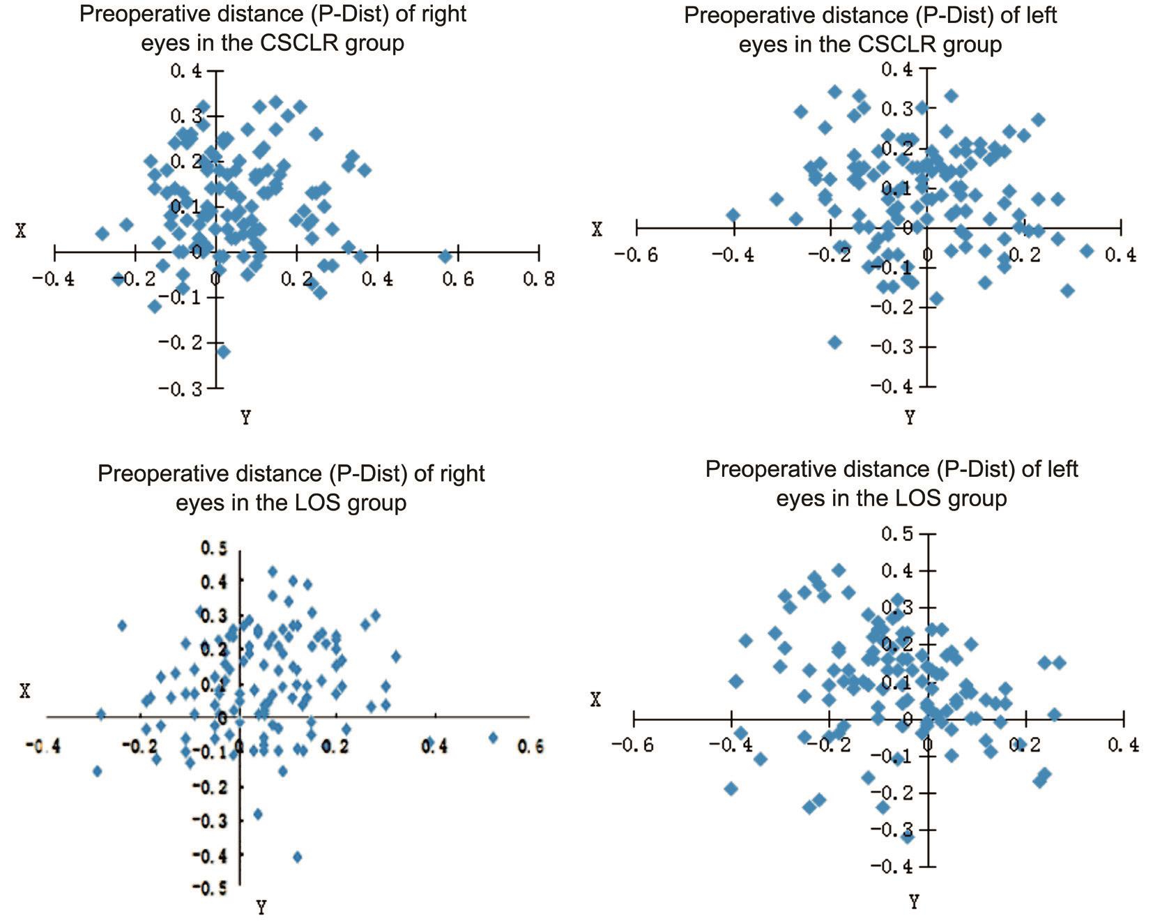
Figure 1 Preoperative scatterplots of the location of the pupil center relative to the visual axis between the CSCLR group and the LOS group.

Figure 2 Change in UDVA in the two groups 6mo postoperatively.
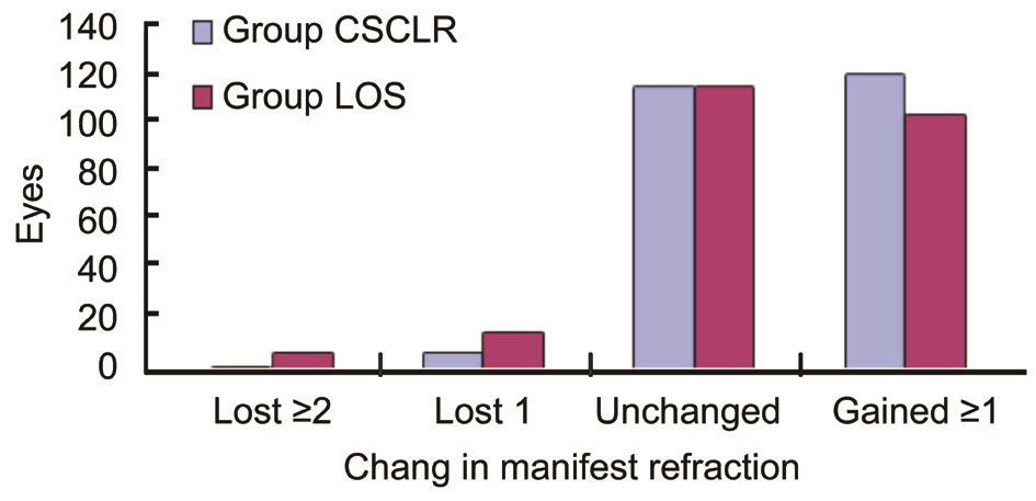
Figure 3 Change in BSCVA in the two groups 6mo postoperatively.
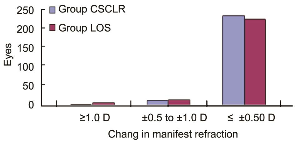
Figure 4 Change in MRSE in the two groups 6mo postoperatively.
DISCUSSION
Over the years, confusion and conflicting definitions over the various axes have been sources of much controversy surrounding the question of what is the appropriate centration technique in corneal refractive surgery. Many axes of the eye can be described, such as the optical axis, pupillary axis, line of sight, visual axis, and line of fi xation. LOS is def i ned by the fi xation point at one end and the center of the entrance pupil at the other[10,12]. The pupillary axis has been described as the line perpendicular to the cornea that passes through the center of the entrance pupil, which also passes through the center of curvature of the corneal surfaces. The angle between the pupillary axis and the LOS is the angle lambda and has been clinically measured to be around 3°-6°[14-15]. Another angle that is frequently described, but is impossible to measure in the eye, is angle kappa, which is the angle between the pupillary axis and the theoretical visual axis[16-17]. The visual axis is defined as the line between the fixation point and the foveabut, in fact, it is diff i cult to locate the visual axis. The current study showed that the CSCLR is the ideal anatomical site,close to this intersection point on the corneal surface[17]. Chan and Boxer Wachler[7] also conf i rmed that the CSCLR was the closest approximation to the visual axis. Additionally, the CSCLR obtained from corneal topography may not accurately determine the location of the visual axis. Recent studies[18-19]have shown differences in the location of the visual axis, as estimated by the CSCLR and the LOS, in a population of myopic refractive surgery candidates, indicating that a precise and optimal def i nition of centration strategy is necessary.
Table 3 Postoperative parameters for both groups

CSCLR: Coaxially sighted corneal light ref l ex; LOS: Line of sight. Statistically signif i cant (Ρ<0.05).
Table 4 Postoperative aberrations changs between the two groups
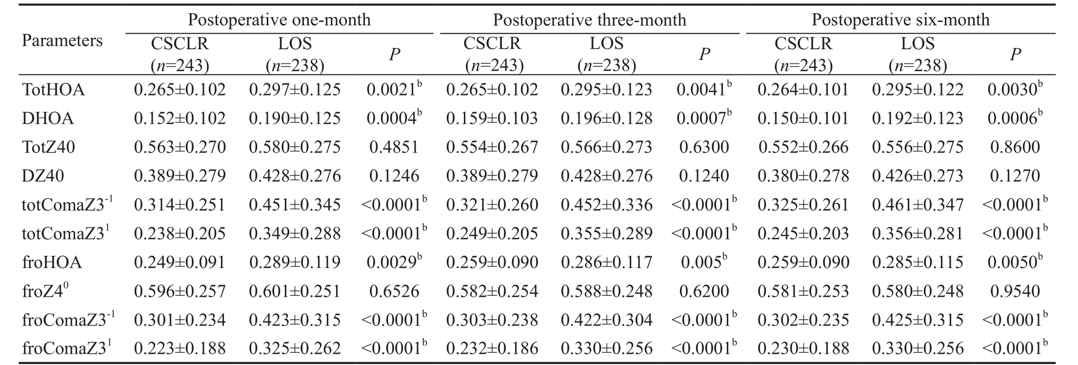
CSCLR: Coaxially sighted corneal light ref l ex; LOS: Line of sight. bStatistically signif i cant difference using the Student’s t-test (Ρ<0.05).
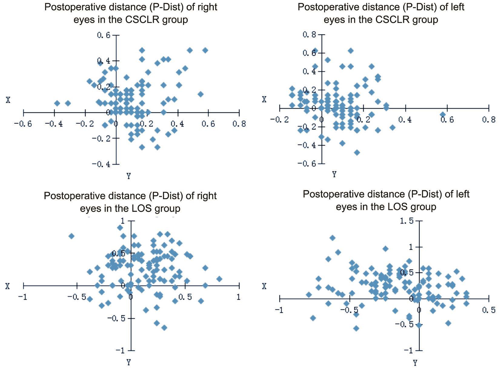
Figure 5 Scatterplots of pupil center shifts relative to the visual axis between the CSCLR and the LOS group postoperatively.
Table 5 Postoperative contrast sensitivity changs between the two groups

CSCLR: Coaxially sighted corneal light reflex; LOS: Line of sight. cStatistically significant difference using the Wilcoxon signed rank test(Ρ<0.05).
A disadvantage of selecting the LOS is the documented change in the pupil center under different light conditions[20-21]. This change can result in relatively large changes in refraction because the curvature of the cornea changes with location.Yang et al[22] reported that they measured the pupil center of 70 eyes in dark and light environments and pharmacologically dilated conditions, and found that when the pupil diameter became larger, the pupil center continued to move the eye temporally, on average by 0.133 mm. Some scholars believe that the pupil center is a virtual image created by the bundle of rays of light across the aqueous humor and cornea refracted into the eye, so its reliability is questionable[23]. Thus, centering on the LOS maybe a greater risk for myopic LASIK. To center on the CSCLR may be preferable because it is not affected by pupil size. If the surgeon sights monocularly, directly behind the fi xation light, the patient’s corneal light ref l ex will appear to be decentered nasally in the pupil; the projection of the corneal light reflex onto the corneal surface will correspondingly be located nasal to the point where the line of sight and the cornea intersect. That is, the corneal light ref l ex will be located nasal to the optimal centration point for corneal surgical procedures.If the LOS is used to guide centration for a patient with a large kappa angle, there would be an error in marking the center.
This study used topographical methods to measure centration of corneal procedures accurately from the pupil, and the surgeon removed the centration to the cornea ref l ex points of the visual axis during the LASIK procedure. The outcomes in this comparison indicated that both CSCLR and LOS centration are safe, accurate, and efficacious. Safety was indicated in this large cohort of 481 eyes by no loss of more than one line of vision. The loss of one line of CDVA falls within measurement variability and is considered clinically insignif i cant. In the CSCLR group, one eye lost more than one line of BSCVA and six eyes lost one line of BSCVA. In the LOS group, six eyes lost more than one line of BSCVA and 14 eyes lost one line of BSCVA. A UCVA of 20/20 or better was achieved in 84% of the CSCLR group and 86% of the LOS group; there was no statistically signif i cant difference between the groups. The refractive outcomes in the current study were not statistically signif i cantly different between the CSCLR and LOS groups. At postoperative 6mo, 95% of eyes in the CSCLR group and 94% of eyes in the LOS group were within ±0.50 D of the intended MRSE. Taken together, these outcomes indicate that a good safety index and an eff i cacy index could be achieved using both ablation strategies. Mrochen et al[3]compared myopic laser treatment, centering on the visual axis versus the line of sight. However, in that study, they performed comparisons on the early (1-month postoperative)data, which may not be accurate because refractive stability may not be achieved. In our study, we present longer term follow-up (6-month) data along with objective measures of the optical quality of the eye and visual quality; we also take comprehensive evaluation into consideration by treating closer to the visual axis rather than the line of sight.
In the current study, the pupil center distribution, angle kappa, ablation center and whole cornea, corneal anterior, and posterior surface of high-order aberrations were measured using a Pentacam, which is based on Scheimpflug imaging,and performed a Zernike analysis for the whole cornea by measuring height data. Using the topographic corneal vertex location has some advantages because this point is reliable and reproducible on topography. Because the CSCLR is the closest approximation to the visual axis, and it represents a stable preferable morphologic reference. Centration on the pupil poses a signif i cant challenge because the pupil center changes with differing illumination and with age[13,24]. If patients possessing larger angle kappa can appear mildly exotropic while fixating on the LOS, it may be result in imprecise alignment of ablation. As this situation, centering on the CSCLR is more benef i cial and desirable strategy, which is not affected by pupil size[19]. The postoperative induction of HOAs in the CSCLR group was statistically signif i cantly lower than in the LOS group. Increased magnitude of coma is indicative of decentration, subclinical or otherwise[21]. Postoperative coma was statistically signif i cantly higher in the LOS group, as the P-distance increased from 0.15 mm to more than 0.25 mm(Ρ<0.05). P-distance increases were less than 0.25 mm in 69.5%of the CSCLR group and in 19.3% in the LOS group; that is,the P-distance increased more than 0.25 mm in 20.6% of the former group and 80.7% of the latter group. The increased coma indicated greater ablation decentration in the LOS group. This outcome was consistent with Reinstein et al[18],who reported that small ablation decentration (P-distance<1 mm) showed no significant difference in UCVA or MRSE postoperatively, but it was a major cause in the induction of postoperative spherical aberrations and coma. Khakshoor et al[21] reported that centration on the CSCLR for myopic patients within high angle κ values may aid in providing better refractive outcomes and vision quality, which was consistent with our results. Our clinical outcomes indicated that there was no statistically signif i cant difference in the increased magnitude of spherical aberrations postoperatively, but that postoperative coma was statistically signif i cantly higher in the LOS group,and the increased magnitude of coma and the eccentric magnitude of the ablation were positively related (r=0.69,Ρ<0.01). Postoperative spherical aberrations were not affected by decentration because the spherical aberrations were radially symmetric. Centration on the CSCLR reduced positioning errors made by the surgeon when estimating the visual axis and decreased the induction of coma postoperatively.
At 1mo postoperatively, the contrast sensitivity function was statistically signif i cantly different at low frequencies between the two groups, but there was no signif i cant difference at any frequency at 6mo postoperatively. This indicated that the ablation decentration was minor and did not lead to poor visual quality in either group. In the early stages of LASIK surgery, a decrease in CSF was related to factors such as corneal edema,irregularity of the corneal surface, light scattering of the corneal layer, central corneal fl attening, ablation decentration,pupil size and optical zone matching, and haze, all of which can reduce the quality of vision. Our results were consistent with previous studies[21,25]; over time, the CSF returned to the preoperative level by 6mo after LASIK surgery.
A limitation of this study is that we did not choose to centrate on the LOS and the CSCLR on different eyes in the same candidate, which could demonstrate the greater strength of centering on the CSCLR. Another drawback is the follow-up of 6mo, which may not be long enough to identify changes that occur over a longer follow-up.
In conclusion, myopic LASIK centered on the CSCLR appears to represent a more preferable method, which is safe, accurate and efficacious, avoiding suboptimal refractive outcomes and induction of HOAs. Ablation centration closer to the CSCLR may achieve the best optical results and improve the postoperative vision quality. It is expected that larger studies with longer follow-up will further demonstrate the safety and efficacy of the procedure and validate the advantage of this centration technique in myopes.
ACKNOWLEDGEMENTS
Foundation: Supported by the Natural Science Foundation of Shanghai Municipal Commission of Health and Family Planning (No.20134230).
Conflicts of Interest: Zhang J, None; Zhang SS, None; Yu Q,None; Lian JC, None.
REFERENCES
1 Bueeler M, Mrochen M, Seiler T. Maximum permissible lateral decentration in aberration-sensing and wavefront-guided corneal ablation.J Cataract Refract Surg 2003;29(2):257-263.
2 Yap EY, Kowal L. Diplopia as a complication of laser in situ keratomileusis surgery. Clin Experiment Ophthalmol 2001;29(4):268-271.
3 Mrochen M, Kaemmerer M, Mierdel P, Seiler T. Increased higher-order optical aberrations after laser refractive surgery: a problem of subclinical decentration. J Cataract Refract Surg 2001;27(3):362-369.
4 Kermani O. Automated visual axis alignment for refractive excimer laser ablation. J Refract Surg 2006;22(9 Suppl):S1089-1092.
5 Kermani O, Oberheide U, Schmiedt K, Gerten G, Bains HS. Outcomes of hyperopic LASIK with the NIDEK NAVEX platform centered on the visual axis or line of sight. J Refract Surg 2009;25(1 Suppl):S98-103.
6 Pande M, Hillman JS. Optical zone centration in keratorefractive surgery. Entrance pupil center, visual axis, coaxially sighted corneal ref l ex,or geometric corneal center? Ophthalmology 1993;100(8):1230-1237.
7 Chan CC, Boxer Wachler BS. Centration analysis of ablation over the coaxial corneal light reflex for hyperopic LASIK. J Refract Surg 2006;22(5):467-471.
8 Arbelaez MC, Vidal C, Arba-Mosquera S. Clinical outcomes of corneal vertex versus central pupil references with aberration-free ablation strategies and LASIK. Invest Ophthalmol Vis Sci 2008;49(12):5287-5294.
9 Okamoto S, Kimura K, Funakura M, Ikeda N, Hiramatsu H, Bains HS.Comparison of wavefront-guided aspheric laser in situ keratomileusis for myopia: coaxially sighted corneal-light-reflex versus line-of-sight centration. J Cataract Refract Surg 2011;37(11):1951-1960.
10 Uozato H, Guyton DL. Centering corneal surgical procedures. Am J Ophthalmol 1987;103(3 Pt1):264-275.
11 Chang DH, Waring GO 4th. The subject-fixated coaxially sighted corneal light reflex: a clinical marker for centration of refractive treatments and devices. Am J Ophthalmol 2014 ;158(5):863-874.
12 Schwiegerling JT. Eye axes and their relevance to alignment of corneal refractive procedures. J Refract Surg 2013;29(8):515-516.
13 Wilson MA, Campbell MC, Simonet P. The Julius F. Neumueller Award in Optics, 1989: change of pupil centration with change of illumination and pupil size. Optom Vis Sci 1992;69(2):129-136.
14 Applegate RA, Thibos LN, Twa MD, Sarver EJ. Importance of fixation, pupil center, and reference axis in ocular wavefront sensing,videokeratography, and retinal image quality. J Cataract Refract Surg 2009;35(1):139-152.
15 Basmak H, Sahin A, Yildirim N, Papakostas TD, Kanellopoulos AJ.Measurement of angle kappa with synoptophore and Orbscan II in a normal population. J Refract Surg 2007;23(5):456-460.
16 Hashemi H, KhabazKhoob M, Yazdani K, Mehravaran S, Jafarzadehpur E,Fotouhi A. Distribution of angle kappa measurements with Orbscan II in a population-based survey. J Refract Surg 2010;26(12):966-971.
17 Park CY, Oh SY, Chuck RS. Measurement of angle kappa and centration in refractive surgery. Curr Opin Ophthalmol 2012;23(4):269-275.18 Reinstein DZ, Archer TJ, Gobbe M. Is topography-guided ablation profile centered on the corneal vertex better than wavefront-guided ablation profile centered on the entrance pupil? J Refract Surg 2012;28(2):139-143.
19 El Danasoury AM, Gamaly TO, Hantera M. Multizone LASIK with peripheral near zone for correction of presbyopia in myopic and hyperopic eyes: 1-year results. J Refract Surg 2009;25(3):296-305.
20 Erdem U, Muftuoglu O, Gundogan FC, Sobaci G, Bayer A. Pupil center shift relative to the coaxially sighted corneal light reflex under natural and pharmacologically dilated conditions. J Refract Surg 2008;24(5):530-538.
21 Khakshoor H, McCaughey MV, Vejdani AH, Daneshvar R, Moshirfar M. Use of angle kappa in myopic photorefractive keratectomy. Clin Ophthalmol 2015;29(9):193-195.
22 Yang Y, Thompson K, Burns SA. Pupil location under mesopic,photopic, and pharmacologically dilated conditions. Invest Ophthalmol Vis Sci 2002;43(7):2508-2512.
23 Schruender SA, Fuchs H, Spasovski S, Dankert A. Intraoperative corneal topography for image registration. J Refract Surg 2002;18(5):S624-629.
24 Moshirfar M, Hoggan RN, Muthappan V. Angle Kappa and its importance in refractive surgery. Oman J Ophthalmol 2013;6(3):151-158.
25 Reinstein DZ, Gobbe M, Archer TJ. Coaxially sighted corneal light reflex versus entrance pupil center centration of moderate to high hyperopic corneal ablations in eyes with small and large angle kappa. J Refract Surg 2013;29(8):518-525.