Dear Editor,
Vogt-Koyanagi-Harada (VKH) disease is a cell-mediated autoimmune syndrome directed against melanocytes.It is considered a multisystem disorder characterized by granulomatous panuveitis often associated with neurologic and cutaneous manifestations. The choroid is the main site of autoimmune inf l ammation in ocular tissues[1].
Here we report the case of a 48-year-old woman with diminished vision that began about 15d earlier. The patient has consented to the submission of this Letter for submission to the journal. Her visual acuity was 0.8 in both eyes. She also suffered from headache and hearing loss, and reported a bout of influenza some days before the onset of diminished vision. The anterior segment examinations of both eyes were unremarkable. Lumbar puncture was not performed. The fundus examination revealed peripapillary bilateral exudative retinal detachments, which are a common cause of visual function impairment in VKH patients. Enhanced depth imaging optical coherence tomography (EDI-OCT) has been used to visualize retinal and choroidal structures in patients affected by VKH disease[2], but to our knowledge there have been no previous attempts to analyze the choroidal and vascular features by OCT angiography alone, which is already being used as a dyeless diagnostic tool in clinical ophthalmology.
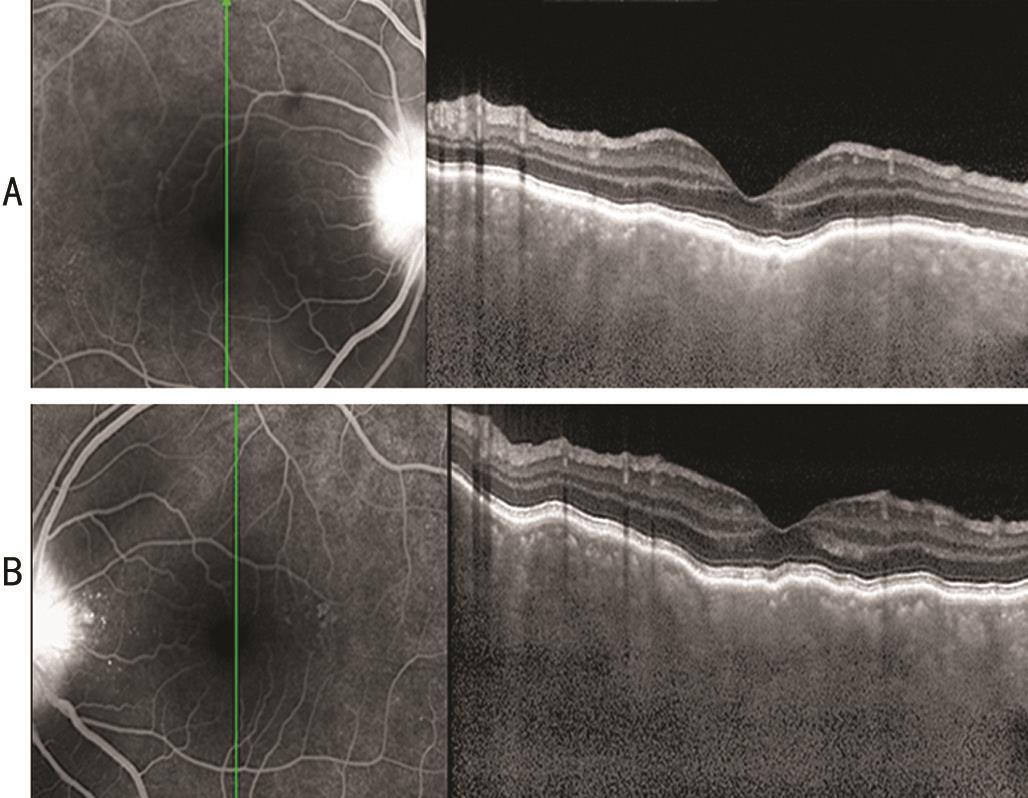
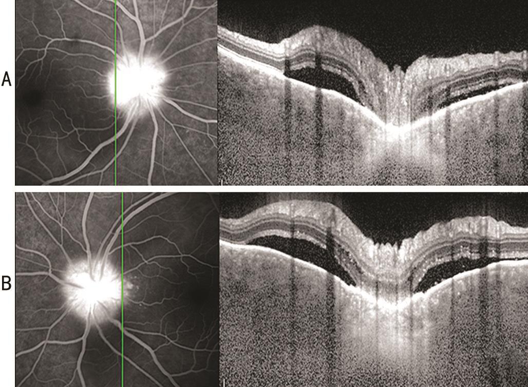
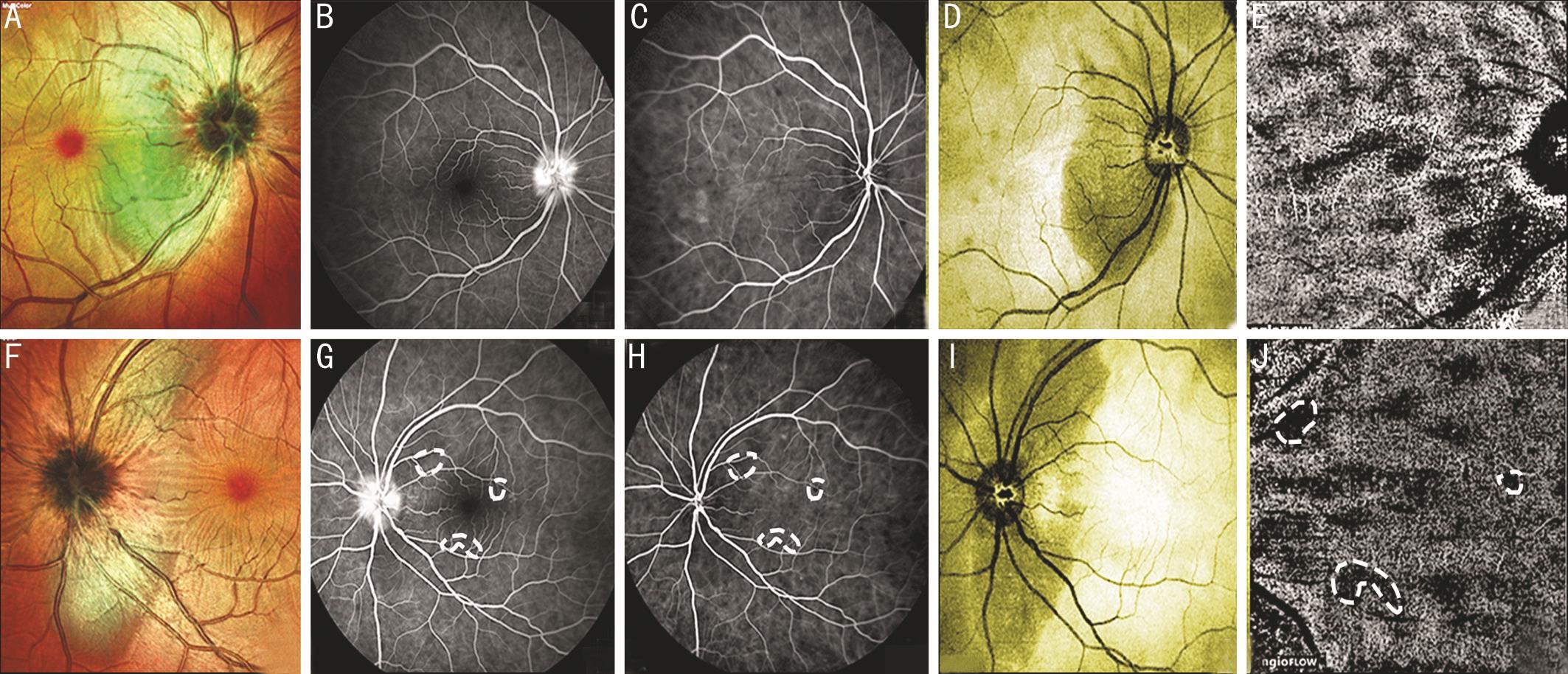
Vertical and horizontal oriented EDI-OCT scans of the macula(Spectralis HRA+OCT Heidelberg Engineering, Heidelberg,Germany) revealed, in both eyes, retinal pigment epithelial(RPE) undulations (Figure 1), peripapillary retinal detachments with fl uid accumulation (Figure 2), a thickened choroid with many hyperref l ective points and choroidal folds.Multicolor fundus imaging revealed an area of sunset glow corresponding to areas of retinal detachment (Figure 3A,3F). Fluorescein angiography showed some hyperreflective perimacular and peripapillary pinpoint leakages in the early phase and optic disk dye staining starting from the earliest phases until the late phases (Figure 3B, 3G), and some hypofluorescence foci. We excluded a diagnosis of acute posterior multifocal placoid epitheliopathy because, the background of this disorder generally includes yellowish well defined circular areas in the fundus that corresponded to the hypofluorescent areas in fluorescein angiography, and these dark areas became hyperf l uorescent in the late phase starting from the edges. Indiocyanine green angiography revealed hypofluorescent spots scattered throughout the fundus from the early phase (Figure 3C, 3H); these spots presumably represent the blockage of dye by inf i ltration of inf l ammatory cells in the choroid and overlying retinal pigment epithelium changes as previously reported[3]. Retinal detachments close to the optic nerve were clearlyl visible as hyporef l ectivey areas on en face scans (Figure 3D, 3I). OCT angiography (Optovue RTVue XR 100 Avanti, Optovue, Inc, Fremont, CA, USA)was normal in the superficial and deep plexus. The outer retinal layer contained irregular hyporef l ective areas, and the choriocapillaris layer showed a mottled background pattern(“patchy”) with speckled areas (Figure 3E, 3J). We performed an 8×8 mm scan of the area. The segmentation was done between 41 micron upper the retinal pigment epithelium and 69 micron underneath the retinal pigment epithelium.To avoid segmentation errors and flow artifact we analyzed corresponding areas on structural en face images and cross sectional OCT b scans, which suggests that the hyporef l ective areas on en face OCT angiography likely represent true patchy areas. These findings confirmed previous study that showed on OCT angiography, multiple dark foci with loss of coriocapillaris that appeared as areas of fl ow void[4].

Figure 1 Vertical macular OCT Spectralis scans exhibiting RPE undulations with choroidal hyperref l ective spots.

Figure 2 Vertical peripapillary OCT Spectralis scans showing typical retinal detachments with accumulation of fl uid.

Figure 3 Multicolour images showing the sunset glow pattern corresponding to areas of detachment (A, F); early phase fl uorescein angiography showing perimacular and peripapillary pinpoint leakages (demarcated by a circle), optic disk hyperfluorescence, and several foci of hypof l uorescence (B, G); early phase green indocyanine angiography showing several hyperf l uorescence points and many small areas of hypof l uorescence (demarcated by a circle), that probably correspond to inf i ltration of inf l ammatory cells in choroidal tissue (C, H); en face scan showing a dark area surrounding the optic disk (D, I); OCT angiography showing a “patchy” pattern in the choriocapillaris layer (demarcated by a circle) (E, J).
In conclusion, OCT angiography could be a useful tool with which to examine vascular alterations in acute VKH disease, particularly because it is non invasive and can be repeated frequently unlike other angiographic tests. However,additional studies are needed to determine if it can represent an alternative to fl uorescein and indiocyanine green angiography in the diagnosis of VKH disease.
ACKNOWLEDGEMENTS
The authors thank Jean Ann Gilder (Scientif i c Communication srl., Naples, Italy) for editing the manuscript.
Conflicts of Interest: Cennamo GL, None; Romano MR,None; Iovino C, None; de Crecchio G, None; Cennamo G,None.
REFERENCES
1 Rao NA. Pathology of Vogt-Koyanagi-Harada disease. Int Ophthalmol 2007;27(2-3):81-85.
2 Tsuboi K, Nakai K, Iwahashi C, Gomi F, Ikuno Y, Nishida K. Analysis of choroidal folds in acute Vogt-Koyanagi-Harada disease using high-penetration optical coherence tomography. Graefes Arch Clin Exp Ophthalmol 2015;253(6):959-964.
3 Agrawal RV, Biswas J, Gunasekaran D. Indocyanine green angiography in posterior uveitis. Indian J Ophthalmol 2013;61(4):148-159.
4 Aggarwal K, Agarwal A, Mahajan S, Invernizzi A, Mandadi SK, Singh R,Bansal R, Dogra MR, Gupta V; OCTA Study Group. The role of optical coherence tomography angiography in the diagnosis and management of acute Vogt-Koyanagi-Harada disease. Ocul Immunol Inf l amm 2016;20:1-12.