INTRODUCTION
Toxocariasis is one of the most common zoonotic infections worldwide[1-2]. The clinical presentation of toxocariasis in humans varies widely, with some patients having an asymptomatic infection and others developing severe organ damage. Symptom severity is dependent upon parasite load, larval migration site, and host inflammatory response.Moreover, systemic toxocariasis (also known as visceral larva migrans) and ocular toxocariasis can occur, which are particularly dependent upon on the involved organ[2-3].Ocular toxocariasis is a clinically well-defined manifestation of an intraocular Toxocara larvae infection. This condition is an important cause of childhood vision loss, with ocular toxocariasis affecting both the retina and optic nerve. The ocular infection can also severely affect vision in adults,particularly in Asian adults because of cultural food habits[2,4].Several case reports of toxocariasis involving the optic nerve have been published[5], but most ocular toxocariasis studies have focused on retinal damage, including retinal granuloma,epiretinal membrane, macular edema, and retinal detachment.Therefore, the clinical features, diagnosis, treatments, and disease courses of Toxocara optic neuropathy remain unclear.The current study evaluates distinguishing clinical features and the course of Toxocara optic neuropathy.
METHODS
All study conduct adhered to the tenets of the Declaration of Helsinki. This study was approved by the Institutional Review Board at Pusan National University Yangsan Hospital(Yangsan, Korea). We retrospectively evaluated Toxocara optic neuropathy between 2008 and 2016. Patients were diagnosed with Toxocara optic neuropathy if all of the following were present or true: 1) acute optic neuropathy [sudden onset of decreased visual acuity or visual field defect with a relative afferent pupillary defect (RAPD)]; 2) positive serum enzymelinked immunosorbent assay (ELISA) titers for Toxocara canis IgG (ELISA titer of >0.250 considered serologically positive based on a previous study showing 92.2% sensitivity and 86.6%specificity at an optical density =0.250)[6]; 3) all other possiblecauses of optic neuropathy ruled out, including ischemic optic neuropathy, retinal vessel occlusion, autoimmune disease,in flammatory conditions, cancer masquerade syndromes, and other infectious etiologies.
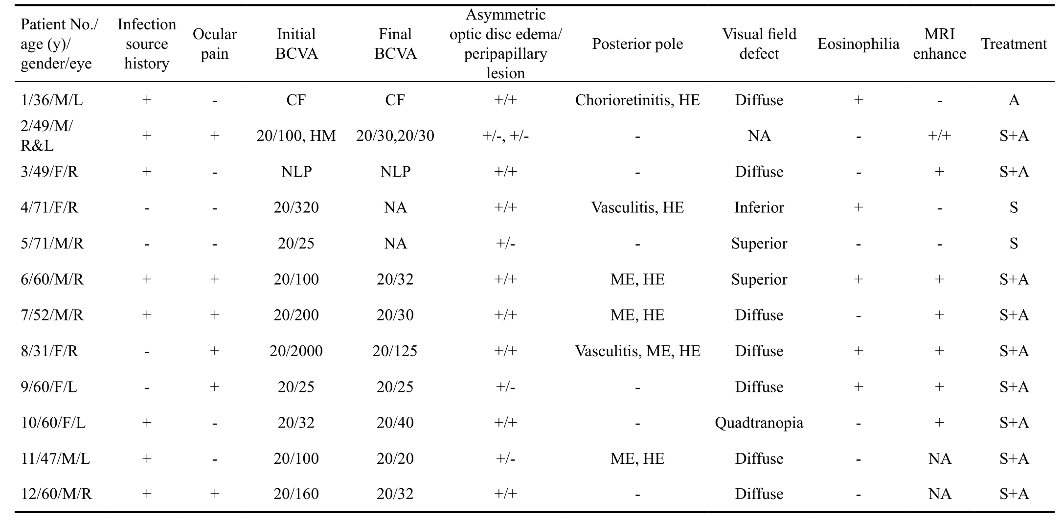
Table 1 Demographics and clinicalfindings of Toxocara optic neuropathy

CF: Countingfingers; HM: Hand movements; NLP: No light perceptions; ME: Macular edema; HE: Hard exudation; NA: Non-applicable;S: Corticosteroid pulse treatment with slow tapering; A: Albedazole. Eosinophil counts >5% (normal range, 1% to 5%).
We investigated possible Toxocara infection sources and conducted a standardized interview, ensuring complete responses and participant comprehension to enhance interview validity. Standard interview questions included queries about ingestion of possible contaminated sources (raw animal liver, meat, and animal blood), history of pet ownership, and occupation-associated contact with animals or soil. Bestcorrected visual acuity (BCVA), intraocular pressure (IOP),slit-lamp biomicroscopyfindings, dilated fundus examination findings, Humphrey visual field testing results, anterior chamber/vitreous inflammatory status were reviewed in all subjects. Optic disc and fundus findings were photographed with a fundus camera (Kowa Co. Ltd., Tokyo, Japan) at each follow-up visit and changes in the optic disc and fundus were evaluated. Cross-sectional images of the optic disc and peripapillary area was obtained and evaluated using spectraldomain optical coherence tomography (SD-OCT; Spectralis,Heidelberg Engineering, Heidelberg, Germany). Subjects were also tested for eosinophilia (defined as >500 eosinophils/µL in peripheral blood or ≥5% of total white blood cell count)and underwent contrast-enhanced orbital magnetic resonance imaging (MRI).
Subjects with Toxocara optic neuropathy were treated with systemic corticosteroid therapy (intravenous steroid pulse therapy for 3d followed by tapering with oral prednisolone for 2wk), anti-parasitic therapy (400 mg albendazole twice a day for 2wk), or a combination of both treatments. Topical corticosteroid therapy (prednisolone acetate 1% four times a day) was also used when anterior chamber in flammation was present.
Treatment outcome was defined as the difference between initial and final visual acuity in patients who had at least 1-month follow-up. Snellen BCVA measurement was converted to the logarithmic of the minimum angle of resolution (logMAR) for all data analyses. Wilcoxon’s rank sum test was used to test statistical significance.
RESULTS AND DISCUSSION
Thirteen eyes of twelve patients were diagnosed with Toxocara optic neuropathy and demographic and clinical features were summarized in Table 1.
Ocular toxocariasis predominantly occurs in men[1,3,7-8].However, our study showed that Toxocara optic neuropathy can also affect women in a similar manner. Most cases included in the current study were unilateral, but one subject did have bilateral involvement. Six subjects (50%) had ocular pain at the time of optic neuropathy presentation and the other six patients had painless acute optic neuropathy. We revealed a strong association between Toxocara optic neuropathy and ingestion of uncooked animal products. Eight patients (66.7%) had identification of a possible infection source, with two patients having a history of puppy/kitten exposure and six patients having history of ingesting raw animal liver, raw meat, or red ants. The remaining four patients (33.3%) had a nonspecific history. This information highlights the importance of asking adult optic neuropathy patients about their consumption of raw meat or liver. Unfortunately, we were not able to elucidate the timing between raw meat or liver ingestion and ocular symptom onset. Previous studies have shown that Toxocara larvae can survive in human tissues for over 10y, therefore,when Toxocara optic neuropathy is suspected, the patient should be asked about raw meat ingestion up to 10y ago[5,9].
However, a definitive diagnosis of ocular toxocariasis can be obtained via histological confirmation of the Toxocara larva or its fragments in infected tissue samples. Collection of intraocular tissue is difficult and rarely warranted in clinical practice. Thus, Toxocara IgG ELISA testing is likely an important diagnostic tool and should be done immediately in patients with suspected Toxocara optic neuropathy.Ahn et al[2]reported that eosinophil was not as helpful as serum anti-Toxocara IgG test in ocular toxocariasis diagnosis(only 11.6% had eosinophilia). However, eosinophilia may indicate the presence of Toxocara larvae, as show in previous reports[1,7], and our study showed 41.6% patient had eosinophilia. Therefore, we recommended that eosinophilia would be additional diagnostic clues for Toxocara optic neuropathy diagnosis if present.
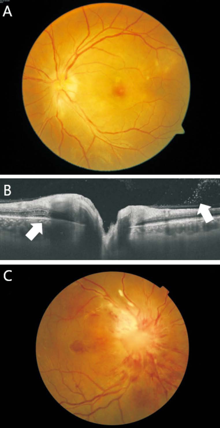
All eyes had optic disc edema; ten eyes had asymmetric and/or sectorial optic disc edema with associated subretinal fluid or peripapillary infiltration (Figure 1A). SD-OCT image of asymmetric optic disc edema with peripapillary lesions showed optic disc edema with subretinal fluid and moderately hyper-reflective mass like lesion, which had posterior shadowing (Figure 1B). The remaining three eyes had diffuse,generalized optic disc edema without peripapillary lesion.Six eyes had retinal lesions with optic disc swelling because of chorioretinitis, retinal vasculitis, macular edema, and hard exudates (Figure 1C). Yang et al[5]published a Toxocara optic neuropathy case series and showed that optic disc swelling was present in all patients, but ranged from subtle to severe.Circumpapillary lesions were presented in all patients and eventually developed retinal lesions[5]. These clinical signs were also present in our Toxocara optic neuropathy patients,we therefore highlight that asymmetric and/or sectorial optic disc edema with peripapillary lesion may be important clinical clue in Toxocara optic neuropathy. Interestingly, the current study showed that Toxocara optic neuropathy can present as a diffuse optic disc swelling without peripapillary and retinal lesions, in addition, some patients had no significant MRI enhancement. These differences in presentation may represent differences in disease severity and/or stage. Disease severity depends not only on the number of larvae ingested,but also on the severity of a patient’s allergic reaction[10-11].As a result, patients with atopy may experience more severe toxocariasis[10-11]. Pathologic manifestations result from the immune-mediated in flammatory response directed against the excretory-secretory larvae antigens. These antigens are released from larvae’s outer epicuticle coat, which readily sloughs off when bound by specific antibodies[12]. These antigens are a mix of glycoproteins, including a potent allergenic component named tubulin alpha chain -1 (TBA-1)[11,13]. Hayashi et al[13]showed that the optic nerve may be a migratory route of Toxocara canis from the brain to the eye in an animal model.Therefore, the optic disc may appear normal while Toxocara canis migrate within the nerve, before development of optic disc edema. As a result, patients suspected of having Toxocara that effects the optic nerve should be closely monitored.

Figure 1 Ocular features of Toxocara optic neuropathy patients
A: Fundus photograph of Case 1 at the initial presentation,asymmetric optic disc edema with peripapillary infiltrating lesion and chorioretinits are apparent; B: Optical coherence tomography crosssectional image of Case 11, optic disc edema with subretinal fluid and vitreous opacity (white arrow) are apparent; C: Fundus photograph of Case 8 at the initial presentation, severe optic disc edema with macular lesions (retinal vasculitis, hard exudates, and severe macular edema) are apparent.
Optic nerve enhancement was observed on orbital MRI in eight of eleven eyes (72.7%) with available MRI. Optic nerve enhancement was localized to the orbital portion of optic nerve in all patients. There were no abnormal findings in the extraorbit portion of optic nerve and brain parenchyma of any patient. Humphrey visual filed testing had been performed in ten patients. Testing results most commonly showed a diffuse visual field defect in eyes with Toxocara optic neuropathy(Table 1).
Nine patients were treated with intravenous pulse corticosteroid therapy and albendazole combination therapy, two patients were treated with intravenous pulse corticosteroid therapy, and one patient was treated with albendazole mono therapy. Eleven eyes of ten patients were included in the comparison between initial and final visual acuity. Visual acuity significantly improved following treatment, mean BCVA was 0.94±0.56 logMAR [range:no light perception (NLP) to 20/20)] at baseline and 0.47±0.59(range: NLP to 20/20) at the final visit (P=0.02). Four eyes(36.4%) had severe vision loss (worse then 20/200) at the initial visit, and then two eyes among four eyes with severe vision loss at initial visit remained severe vision loss at thefinal visit. Treatment of toxocariasis has been standardized with steroid treatment protocols, but efficacy of anti-helminthics has not been established[14]. Nevertheless, we could not compare treatment outcomes after different treatment regimen,Ahn et al[2]reported that albendazole and oral prednisolone combination therapy significantly lowered the 6-month recurrence rate compared with corticosteroid monotherapy. We therefore recommend combined corticosteroid and albendazole treatment may effective for reducing optic nerve in flammation and recurrence.
Our study had several limitations. First, subject interviews were based on standardized questionnaires administered at the time of initial patient presentation. Using careful documentation, we had to rely on patient interviews to identify probable Toxocara infection sources. Second, treatment outcomes were not examined by treatment method and it is possible that visual outcomes may differ for each treatment type (corticosteroid, anti-helminthics, and combination therapy).Further prospective, randomized trials are needed to compare treatment outcomes among the various treatment protocols.
In conclusion, asymmetric optic disc swelling in the presence of a peripapillary and/or retinal lesion, and a history of raw meat ingestion were important clinical findings in adult patients with Toxocara optic neuropathy. Additionally,immediate confirmation of Toxocara using IgG ELISA testing and measuring eosinophil count may be helpful for early diagnosis of Toxocara optic neuropathy. Corticosteroid and albendazole combination therapy would help to prevent serious visual impairment in patients with Toxocara optic neuropathy.
ACKNOWLEDGEMENTS
conflicts of Interest:Choi KD, None; Choi JH, None; Choi SY, None; Jung JH, None.
REFERENCES
1 Rubinsky-Elefant G, Hirata CE, Yamamoto JH, Ferreira MU. Human toxocariasis: diagnosis, worldwide seroprevalences and clinical expression of the systemic and ocular forms. Ann Trop Med Parasitol 2010;104(1):3-23.
2 Ahn SJ, Woo SJ, Jin Y, Chang YS, Kim TW, Ahn J, Heo JW, Yu HG,Chung H, Park KH, Hong ST. Clinical features and course of ocular toxocariasis in adults. PLoS Negl Trop Dis 2014;8(6):e2938.
3 Stewart JM, Cubillan LD, Cunningham ET. Prevalence, clinical features,and causes of vision loss among patients with ocular toxocariasis. Retina 2005;25(8):1005-1013.
4 Choi D, Lim JH, Choi DC, Paik SW, Kim SH, Huh S. Toxocariasis and ingestion of raw cow liver in patients with eosinophilia. Korean J Parasitol 2008;46(3):139-143.
5 Yang HK, Woo SJ, Hwang JM. Toxocara optic neuropathy after ingestion of raw meat products. Optom Vis Sci 2014;91(11):e267-e273.
6 Jin Y, Shen C, Huh S, Sohn WM, Choi MH, Hong ST. Serodiagnosis of toxocariasis by ELISA using crude antigen of Toxocara canis larvae.Korean J Parasitol 2013;51(4):433-439.
7 Smith H, Holland C, Taylor M, Magnaval JF, Schantz P, Maizels R. How common is human toxocariasis? Towards standardizing our knowledge. Trends Parasitol 2009;25(4):182-188.
8 Woodhall D, Starr MC, Montgomery SP, Jones JL, Lum F, Read RW, Moorthy RS. Ocular toxocariasis: epidemiologic, anatomic, and therapeutic variations based on a survey of ophthalmic subspecialists.Ophthalmology 2012;119(6):1211-1217.
9 Girdwood RW, Smith HV, Bruce RG, Quinn R. Human Toxocara infection in west of Scotland. Lancet 1978;1(8077):1318.
10 Teo CG, Singh M, Ting WC, Ho LC, Ong YW, Seet LC. Evaluation of the common conditions associated with eosinophilia. J Clin Pathol 1985;38(3):305-308.
11 Yahiro S, Cain G, Butler JE. Identification, characterization and expression of Toxocara canis nematode polyprotein allergen TBA-1.Parasite Immunol 1998;20(8):351-357.
12 Iddawela RD, Rajapakse RP, Perera NA, Agatsuma T. Characterization of a Toxocara canis species-specific excretory-secretory antigen (TcES-57)and development of a double sandwich ELISA for diagnosis of visceral larva migrans. Korean J Parasitol 2007;45(1):19-26.
13 Hayashi E, Akao N, Fujita K. Evidence for the involvement of the optic nerve as a migration route for larvae in ocular toxocariasis of Mongolian gerbils. J Helminthol 2003;77(4):311-315.
14 Ahn SJ, Ryoo NK, Woo SJ. Ocular toxocariasis: clinical features,diagnosis, treatment, and prevention. Asia Pac Allergy 2014;4(3):134-141.