INTRODUCTION
Cataract surgery has become less invasive and more precise, and maximized postoperative visual acuity is a primary concern to meet the higher expectations of patients.Patients with cataract and corneal astigmatism today undergo cataract surgery with the hope of an emmetropic eye, so both the spherical and cylindrical values of the refractive error should be addressed. Obtaining accurate measurements of corneal astigmatism is a mandatory step for the planning of toric intraocular lens (IOLs) implantation. However, corneal astigmatism has been traditionally computed based on only the anterior corneal measurement and using the standardized corneal refractive index of 1.3375, with the presumptive constant ratio between the anterior and posterior corneal surface[1]. A neglect of posterior corneal measurement may result in incorrect estimation of the total corneal astigmatism.Calculations based on the total corneal astigmatism rather than the keratometric astigmatism reduce the residual refractive astigmatism after surgery[2]. Actual measurements of the posterior corneal curvature for evaluations prior to cataract surgery may reduce the amount of unexpected postoperative refractive astigmatism compared to keratometric astigmatism based on anterior corneal astigmatism alone[3].
Topography is used in corneal imaging with devices such as the Orbscan and Scheimpflug-based technology (e.g. the Pentacam). The topography devices share the advantage of being able to provide posterior corneal toricity. In most studies, the total corneal astigmatism was measured by the devices adopting the algorithm of a vector summation of the astigmatism on the two corneal surfaces with the assumption that the rays refract to the posterior surface was paralleled and corneal thickness could be neglected[4-9].
The Cassini color light emitting diode (LED) corneal analyzer(Cassini, i-Optics, the Hague, the Netherlands) is a neoteric topographer adopting a nonrotationally symmetric projection pattern. The device emits up to 700 distinct LED spots with 3 colors, which project straight on corneal surface. Then the captured refracted picture is analyzed and an elevationbased corneal topography is provided[10]. The acquisition is instantaneous, preserving more robust tear film and managing eye movement independent measurement as the algorithm does centroid detection. It measures the reflection of the second Purkinje images to reconstruct the posterior surface and utilize ray tracing technology to compute the total corneal astigmatism. Cassini doesn’t use monograms nor vector analysis for this calculation, but actually customized ray traced wavefront aberrations reconstructions.
The total corneal astigmatism has been investigated to a very limited extent in the Chinese population, but it is important to obtain an astigmatism calculation based on the correct reconstruction of the true anterior and posterior data without assuming the corneal thickness in order to optimize premium IOL choice and to obtain desired post-operative outcomes. The aim of this study was to explore the effect of posterior corneal astigmatism on the total corneal astigmatism and the error caused by substituting the simulated keratometric (simulated K)corneal astigmatism for the total corneal astigmatism in agerelated cataract population using the Cassini color LED corneal analyzer.
SUBJECTS AND METHODS
A total of 164 patients (71 men and 93 women; 211 eyes) on the waiting list for cataract surgery at Tianjin Eye Hospital(Tianjin, China) who underwent corneal power measurements with the Cassini color LED corneal analyzer (Cassini, i-optics)from April 2015 to August 2015 were included in the study.The study adhered to the tenets of the Declaration of Helsinki and was approved by the Ethics Committee of Tianjin Eye Hospital. All of the subjects provided informed consent for participation in the study. The inclusion criteria were patients with age-related cataract and those with good-quality Cassini scans (overall quality factor ≥85% and posterior quality ≥90%,with a green mark presented). The exclusion criteria were an irregular astigmatism, contact lenses wearing, previous ocular trauma or ocular surgery, corneal disease and a history of ocular inflammation. The patients were divided into two groups, a younger group and an elder group, based on a critical age of 60y to evaluate the effects of aging.
Preoperative corneal measurements were performed on the subjects by Cassini. A scan was qualified for the analysis when all of the quality factors presented a green mark (with a quality factor greater than 85%). Otherwise, the scan was repeated. All of the measurements were obtained by the same experienced technician. Four categories of astigmatic values were obtained as follows: 1) keratometric astigmatism (astigmatism of the simulated K reading): corneal astigmatism from the anterior corneal measurement, which is based only on the assumed constant ratio between the anterior and posterior surfaces using Gullstrand’s definition. The real refractive index has been replaced by the pseudo index of 1.3375, which we call the “keratometric index”; 2) anterior astigmatism: astigmatism from the anterior corneal surfaces over the 3 mm zone, which is based upon the real refractive index (1.376 for the corneal tissue); 3) posterior astigmatism: astigmatism derives from the posterior surface reconstructed on the basis of the reflection of the second Purkinje images. It is calculated based on a point reflection ray tracing measurement and the real indices of refracting media (1.376 for the corneal tissue and 1.336 for aqueous humor); 4) total corneal astigmatism: astigmatism based on the reconstruction of both anterior and posterior corneal surfaces by ray tracing technology and a specific algorithm over the central 3 mm zone, which derives from the wavefront aberration and takes into account the effect of the corneal thickness.
We classified the astigmatism as “with the rule (WTR)” when the meridian of the maximal convergent power was within 90°±30°. In contrast, the astigmatism was classified as “against the rule (ATR)” when the meridian of the maximal convergent power was within 0°±30°. Otherwise, the astigmatism was classified as oblique.
The power vector method proposed by Thibos et al[11] was adopted for the analysis. The astigmatism was converted into J0 (power of Jackson cross cylinder at 90° and 180°) and J45(power of Jackson cross cylinder at 45° and 135°) values using the following formulae:
where C is the negative cylinder power and α is the cylinder axis. A positive J0 value indicates a WTR astigmatism and a negative J0 value indicates an ATR astigmatism. Absiluto meridian difference (AMDR) was defined as the absolute meridian difference between the anterior and posterior corneal astigmatisms and was calculated as the absolute value of the difference of the cylinder axis.
To describe the effect of posterior astigmatism on the magnitude of the total corneal astigmatism and the estimation error caused by the use of the astigmatic magnitude of the simulated K instead of that of the total cornea, we calculated the magnitude differenceSimK-Tca (magnitude differenceSimK-Tca is magnitude of simulated K minus magnitude of total cornea).Then, this difference was compared between the age groups and anterior astigmatism types. To compare the astigmatisms of the simulated K and total cornea both in magnitude and axial orientation, we drew double-angle plots and calculated the vector difference between the two measures using vector analysis[12-13]. The correlation between the anterior astigmatism,the posterior astigmatism and the total corneal astigmatism was evaluated for the J0 and J45 components.
Table 2 The distribution of the patients based on astigmatism type

ATR: Against the rule; WTR: With the rule.
Table 1 A summary of the patient demographics

Table 3 A summary of the 4 calculations of the corneal astigmatism for the whole patient population

SimK: Simulated keratometry.
Statistical Analysis Pearson’s correlation analyses were used for parametric parameters, while Spearman’s correlation analyses were used for non-parametric parameters. The Wilcoxon signed-rank test was used to compare the magnitude of the simulated K and total corneal astigmatism, while the Mann-Whitney U test and Kruskal-Wallis test were used to compare the magnitude and vector components of each measure of astigmatism among the different groups. Linear regression analyses were performed to assess the relationship between the J0 and J45 components of the anterior astigmatism,the posterior astigmatism and the total corneal astigmatism and the magnitude differenceSimK-Tca and anterior corneal J0, AMDR.The Chi-square test was used to compare the negative and positive proportions of the magnitude differenceSimK-Tca between the age groups and the magnitude differenceSimK-Tca between the different anterior astigmatism types. A corrective regression formula was used to analyze the relationship between the magnitude of the simulated K and total corneal astigmatism for which the constant was not included: magnitude of total cornea=β×magnitude of the simulated K, where β indicates the magnitude of the adjustment made to the magnitude of simulated K to approximate that of the total cornea.
All the statistical analyses were managed by SPSS version 21.0(SPSS, Inc., USA). A difference was considered statistically significant when the P value was smaller than 0.05.
RESULTS
This study included a total of 211 eyes from 164 patients. The average age of the patients was 66.8±9.0y (range: 45-83y).The patients were divided into 2 subgroups based on the age:group A, which included middle-aged people younger than 60 years old, and group B, which included elderly people older than 60 years old (Table 1). Group B presented with a higher percentage of ATR astigmatisms. Table 2 shows the distribution of the astigmatism types over the two groups.
The magnitude, vector components, cylinder axis and vector mean of the 4 calculations of the corneal astigmatism are displayed in Table 3. The magnitude of the total cornea was significantly larger than that of the simulated K (1.05±0.63 D vs 0.96±0.57 D, P<0.001, Wilcoxon signed-rank test). The flat meridian of the total corneal astigmatism differed significantly from that of the keratometric astigmatism (86°±41° vs 93°±45°, P=0.001, Wilcoxon signed-rank test). Totally 30.0%of the eyes had a meridian difference of the two calculations larger than 10° and 46.2% of the eyes had a meridian difference larger than 5°.
The cylinder axial orientation of the anterior cornea was horizontal (classified as WTR astigmatism) in 28.0% of the eyes and was vertically aligned (classified as ATR astigmatism)in 59.2% of the eyes. The flat meridian of the posterior cornea was horizontally oriented in 58.3% of the eyes and was vertically aligned in 19.4% of the eyes (Figure 1). This indicated that most of the eyes presented an ATR astigmatism in both the anterior and posterior corneal surfaces because of the negative refractive power on posterior corneal surface.
The averaged magnitude of the posterior corneal surface was 0.34±0.31 D with a mean flat meridian of 96°±66° and a mean vector astigmatism of 0.15 D at 13°. There was no significant difference in the astigmatic magnitude of the 2 age groups (P=0.59, Mann-Whitney test). However, the J0 and J45 components of the posterior astigmatism was slightly different between the two groups, as the younger group showed a value of 0.094 D and the older group showed a value of 0.057 D (P=0.041,Mann-Whitney test). The posterior astigmatic magnitude in the eyes with WTR astigmatism was significantly higher than that in those with ATR astigmatism (0.41 D vs 0.31 D). Meanwhile,in the eyes with oblique astigmatism, the mean power was 0.32 D(P=0.001, Kruskal-Wallis test).

Figure 1 A correlation plot of the flat meridian values of posterior and anterior corneal astigmatisms.
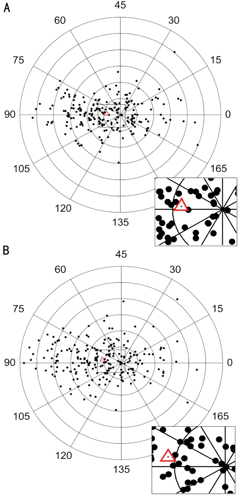
The aggregate mean keratometric astigmatism was 0.41±0.58 D at 88°, while the aggregate mean total corneal astigmatism was 0.57±0.61 D at 85° (Figure 2). The mean vector difference between the two calculations was 0.18 D at 78° (Figure 3).Specifically, the mean vector difference was 0.18±0.26 D at 163° in the ATR astigmatism, 0.20±0.21 D at 177° in the WTR astigmatism, and 0.21±0.23 D at 168° in the oblique astigmatism (Table 4).
Table 5 shows the correlation between the J0 and J45 components of the anterior surface, posterior surface and total cornea. In terms of the J0 component, the posterior corneal astigmatism was positively correlated with the anterior corneal astigmatism and a significant positive correlation was found between the total corneal and the anterior corneal astigmatism (Figure 4).In the younger group A, 43.4% of the eyes had a positive magnitude differenceSimK-Tca value and 56.6% of the eyes had a negative value. Meanwhile, in the elder group B, 27.2% of the eyes had a positive value and 72.8% of the eyes had a negative value (P=0.023; χ2 test). The magnitude differenceSimK-Tca tended to be smaller in the elder group, which suggests the compensatory effect of the posterior surface decreasing with an aging effect. Of the eyes with a positive anterior J0 value,65.7% had a positive magnitude differenceSimK-Tca value and 34.3% had a negative value. Meanwhile, of the eyes with a negative anterior J0 value, 14.2% had a positive value and 85.8% had a negative value (P<0.001; χ2 test), which suggested that the compensatory effect of the posterior surface was more obvious in the eyes with a WTR anterior astigmatism.

Figure 2 Double-angle plots of astigmatism of the simulated keratometry (A) and that of the total cornea (B) Each ring equals 0.5 D, and the mean aggregate astigmatism is signified with a triangle.

Figure 3 Double-angle plots of the vector difference between the astigmatism of simulated keratometry and that of the total cornea Each ring equals 0.5 D, and the mean aggregate vector difference is signified with a triangle.
Table 4 The vector mean of the astigmatism and vector difference between astigmatisms of the SimK and those of the total cornea in different types of astigmatism

ATR: Against the rule; WTR: With the rule; SimK: Simulated keratometry.

Figure 4 The results of the linear regression analysis of the relationship between the J0 components of the posterior cornea and anterior cornea (A) and between those of the total cornea and anterior cornea (B) A: y=0.09+0.11x, R2=0.194, P<0.001; B: y=-0.09+0.9x, R2=0.910,P<0.001.
Table 5 A summary of the correlation between anterior, posterior and total corneal astigmatisms

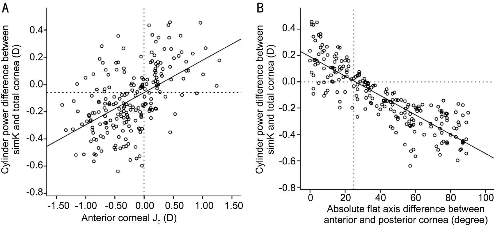
The magnitude differenceSimK-Tca was positively correlated with the anterior corneal J0 (Spearman’s rho=-0.539; P<0.001)and negatively correlated with the AMDR (Spearman’s rho=-0.875; P<0.001). When the anterior corneal J0 was greater than 0.25 D or the AMDR was less than 25°, the magnitude differenceSimK-Tca tended to be a positive value, which indicated an overestimation of total corneal astigmatism by karatometric astigmatism and when the anterior corneal J0 was less than 0.25 D or the AMDR was greater than 25°, the magnitude differenceSimK-Tca demonstrated the opposite trend (Figure 5).When the anterior J0 value was greater than 1.3 D or less than-0.8 D, the errors caused by determining the total corneal astigmatism with the karatometric calculation tended to be greater than 0.25 D.
On the basis of our corrective regression formula, the correlation coefficient β of the magnitude of the total cornea and simulated K was 0.91 in eyes with a positive anterior J0 value and 1.16 in eyes with negative anterior J0 value (Figure 6). This suggests that an underestimation by 16% was indicated in the eyes with an ATR anterior astigmatism and an overestimation by 9% was indicated in the eyes with a WTR anterior astigmatism with the measurement of the keratometric astigmatism.
DISCUSSION
Posterior corneal surfaces contributes to the total corneal astigmatism as well as anterior surface. With a variety of technologies such as Purkinje imaging, scanning-slit topography,and rotating Scheimp flug imaging, previous studies[4-9,14] have reported that the posterior corneal surface had a cylinder power averaged at -0.26 D to -0.78 D. In the present study,the average posterior cylinder power was -0.34 D, ranging from -0.02 D to -0.99 D, which is similar to those of previous reports[4-9,14].
The ray tracing technology of the Cassini traces the refraction of the rays though the anterior corneal surface to the posterior surface and reconstructs both surfaces to accurately calculate the total corneal astigmatism. The repeatability of the magnitude and axis of astigmatism of the Cassini has been evaluated in previous studies, and this new corneal topography device appears to provide a pinpoint accuracy[10,15-17]. It is reported that the corneal power and cylinder measurements obtained outcomes comparative to those of frequently used devices based on Placido rings, Scheimpflug images and monochromatic LEDs[17].

Figure 5 The results of the linear regression analysis of the correlation between the magnitude difference between the simulated K and total cornea and anterior corneal J0 (A) and that of the absolute flat meridian difference between the anterior and posterior surfaces (B)A: y=-0.06+0.24x, R2=0.274, P<0.001; B: y=0.19-0.007x, R2=0.720, P<0.001.

Figure 6 The relationship between the magnitude of simulated K and total corneal astigmatism in eyes with an anterior J0 greater than 0(A) and in eyes with an anterior J0 less than 0 (B) A: y=0.91x, R2=0.959, P<0.001; B: y=1.16x, R2=0.977, P<0.001.
Ignoring the posterior astigmatism may lead to an underestimation of the ATR astigmatism and an overestimation of the WTR astigmatism because most of the posterior surface has a WTR shape and contributes to the ATR astigmatic power. This leads to an undercorrection for the ATR astigmatism and an overcorrection for the WTR astigmatism[14,18-20]. If the errors caused by calculating the total corneal astigmatism from the karatometric measurement could be predictive, this could benefit many clinicians when posterior measurement was out of reach.
In this study, we assessed the effect of the axial orientation difference between the anterior and posterior corneal surfaces on the evaluation of the total corneal astigmatism using the karatometric astigmatism measurement. We determined that the keratometric astigmatism tended to be smaller compared to the total corneal astigmatism as the angle between the cylinder axes of the two surfaces increased. In eyes with a WTR astigmatism on the anterior corneal surface, the presence of an ATR astigmatism on the posterior surface compensated for the corneal astigmatism and thus reduced the total corneal astigmatism. However, in eyes with an ATR astigmatism on the anterior corneal surface, the total corneal astigmatism was aggravated. Therefore, when the anterior J0 was a positive value, the magnitude of keratometric astigmatism tended to be larger than that of the total corneal astigmatism, while when the anterior J0 was a negative value, the opposite tendency was observed. These results reaffirmed the findings that were reported previously[14,21].
We observed no correlation between the astigmatic magnitude of the anterior and posterior corneal surfaces (r=0.075;P=0.277). However, the vector components of the two surfaces were highly correlated (Table 3). This finding was different from those of previous studies[9,21] because most of the eyes in these studies displayed a WTR anterior astigmatism,while most of the eyes in our study showed an ATR anterior astigmatism. A moderate positive correlation between the astigmatic magnitude of the anterior and posterior surfaces was found when the cylinder axis of the anterior cornea oriented horizontally, while no correlation was observed in eyes with anterior cylinder axis oriented vertically[14,22], which was often the case with most cataract patients. Additionally, we observed no correlation between the astigmatic magnitude of the total cornea and posterior corneal surface (r=0.157; P=0.022).
According to the regression model of the relationship between the magnitude differenceSimK-Tca and anterior J0 in eyes with an anterior corneal J0 larger than 1.3 D or smaller than -0.8 D, the anterior astigmatism measurement causes an error of more than 0.25 D. That is, when we address a WTR anterior astigmatism of more than 2.6 D or an ATR astigmatism of more than 1.6 D,the anterior measurement can cause errors that may have an influence on the toric IOL decision. And more remarkable, the magnitude and the axis of posterior corneal astigmatism are not constant and need to be measured precisely particularly for eyes with high ATR astigmatism on the anterior corneal surface because on this occasion, toric IOL implantation would become the first choice for many people. In our study, eyes with an ATR astigmatism larger than 1.6 D accounted for more than 1/4 of the eyes with an ATR astigmatism, so an individual evaluation of the posterior corneal astigmatism in these patients is very necessary.
If the posterior corneal measurements are not available, we recommend a corrective regression formula adjusting the magnitude of keratometric astigmatism to conclude to the total corneal astigmatism, especially in eyes with an anterior corneal J0 out of the range from -0.8 D to 1.3 D. Based on our regression formula, a 9% reduction in the magnitude of keratometric astigmatism is suggested in eyes with WTR astigmatism and a 16% addition is suggested in eyes with ATR anterior astigmatism. Prediction errors in the WTR astigmatism were greater than those of the ATR astigmatism, as Koch et al[23] reported for 5 devices that in the WTR eyes, there were significant WTR prediction errors (0.5 D to 0.6 D) and in ATR eyes, the WTR prediction errors were 0.2 D to 0.3 D.
The magnitude of the keratometric astigmatism tends to be larger than that of the total corneal astigmatism in the elder group. Therefore, the compensatory effect of the posterior surface on the total corneal astigmatism decreases with an advancing age and even turns into an augmentative effect.This confirms the results of the previous studies by Koch et al[14]and Ho et al[24]. The anterior corneal flat meridian shifts from a horizontal position to a vertical position, whereas the posterior corneal flat meridian changes slightly[14,25]. The posterior corneal astigmatism often partly compensates for the corneal astigmatism in young individuals and is often additive to the corneal astigmatism in older people. The total corneal astigmatism estimation relying on anterior cornea measurement does not take into consideration the different effects with age. Therefore, a nomogram based on the anterior measurement customized for younger people would not adapt for that of older people. This may be the reason why the adjustive coefficient of the keratometric astigmatism for the total corneal astigmatism in the present study differs from that of a previous study with a younger population of participants,which recommended a 12% reduction in the magnitude of the keratometric astigmatism in eyes with a WTR astigmatism and a 20% addition of the magnitude of the keratometric astigmatism in eyes with an ATR astigmatism[21].
Limitations of this study can be described as follows: 1)Some patients had their both eyes included for the purpose of having a larger sample size, but the sample size of this study was still small; 2) The average age of the patients in the study was 64 years old, ranging from 45 to 83y. Therefore, agerelated changes could not be fully revealed and conclusions of this study may not be adaptive for application to young patients; 3) The corneal topographer that we used in this study was introduced to only a very confined limit, and prospective studies are required to validate the precision of these posterior corneal measurements.
In summary, to achieve more accurate corneal astigmatism measurements for toric IOL planning, the posterior corneal astigmatism should be valued, especially when we correct for a high ATR astigmatism of the anterior corneal surface. A 9%reduction in the magnitude of the keratometric astigmatism is suggested in eyes with a WTR astigmatism, and a 16%addition is suggested in eyes with an ATR astigmatism if the posterior measurements are not available.
ACKNOWLEDGEMENTS
Foundations: Supported by the National Natural Science Foundation of China (No.81670837); the Scientific and Technological Project of Tianjin Health Bureau(No.2015KY38).
Conflicts of Interest: Jin YY, None; Zhou Z, None; Yuan XY, None; Song H, None; Tang X, None.
REFERENCES
1 Olsen T. On the calculation of power from curvature of the cornea. Br J Ophthalmol 1986;70(2):152-154.
2 Savini G, Næser K. An analysis of the factors influencing the residual refractive astigmatism after cataract surgery with toric intraocular lenses.Invest Ophthalmol Vis Sci 2015;56(2):827-835.
3 Sano M, Hiraoka T, Ueno Y, Itagaki H, Ogami T, Oshika T. Influence of posterior corneal astigmatism on postoperative refractive astigmatism in pseudophakic eyes after cataract surgery. BMC Ophthalmol 2016;16(1):212.
4 Royston JM, Dunne MC, Barnes DA. Measurement of posterior corneal surface toricity. Optom Vis Sci 1990;67(10):757-763.
5 Dunne MC, Royston JM, Barnes DA. Posterior corneal surface toricity and total corneal astigmatism. Optom Vis Sci 1991;68(9):708-710.
6 Prisant O, Hoang-Xuan T, Proano C, Hernandez E, Awwad ST, Azar DT. Vector summation of anterior and posterior corneal topographical astigmatism. J Cataract Refract Surg 2002;28(9):1636-1643.
7 Modis L Jr, Langenbucher A, Seitz B. Evaluation of normal corneas using the scanning-slit topography/pachymetry system. Cornea 2004;23(7):689-694.
8 Dubbelman M, Sicam VA, Van der Heijde GL. The shape of the anterior and posterior surface of the aging human cornea. Vision Res 2006;46(6-7):993-1001.
9 Ho JD, Tsai CY, Liou SW. Accuracy of corneal astigmatism estimation by neglecting the posterior corneal surface measurement. Am J Ophthalmol 2009;147(5):788-795,795.e1-2.
10 Kanellopoulos AJ, Asimellis G. Distribution and repeatability of corneal astigmatism measurements (magnitude and axis) evaluated with color light emitting diode reflection topography. Cornea 2015;34(8):937-944.
11 Thibos LN, Wheeler W, Horner D. Power vectors: an application of Fourier analysis to the description and statistical analysis of refractive error. Optom Vis Sci 1997;74(6):367-375.
12 Holladay JT, Moran JR, Kezirian GM. Analysis of aggregate surgically induced refractive change, prediction error, and intraocular astigmatism. J Cataract Refract Surg 2001;27(1):61-79.
13 Ho JD, Tsai CY, Tsai RJ, Kuo LL, Tsai IL, Liou SW. Validity of the keratometric index: evaluation by the Pentacam rotating Scheimpflug camera. J Cataract Refract Surg 2008;34(1):137-145.
14 Koch DD, Ali SF, Weikert MP, Shirayama M, Jenkins R, Wang L.Contribution of posterior corneal astigmatism to total corneal astigmatism.J Cataract Refract Surg 2012;38(12):2080-2087.
15 Hidalgo IR, Rozema JJ, Dhubhghaill SN, Zakaria N, Koppen C,Tassignon MJ. Repeatability and inter-device agreement for three different methods of keratometry: Placido, Scheimpflug, and color LED corneal topography. J Refract Surg 2015;31(3):176-181.
16 Kanellopoulos AJ, Asimellis G. Color light-emitting diode reflection topography: validation of keratometric repeatability in a large sample of wide cylindrical-range corneas. Clin Ophthalmol 2015;9:245-252.
17 Klijn S, Reus NJ, Sicam VA. Evaluation of keratometry with a novel Color-LED corneal topographer. J Refract Surg 2015;31(4):249-256.
18 Savini G, Versaci F, Vestri G, Ducoli P, Næser K. Influence of posterior corneal astigmatism on total corneal astigmatism in eyes with moderate to high astigmatism. J Cataract Refract Surg 2014;40(10):1645-1653.
19 Tonn B, Klaproth OK, Kohnen T. Anterior surface-based keratometry compared with Scheimp flug tomography-based total corneal astigmatism.Invest Ophthalmol Vis Sci 2014;56(1):291-298.
20 Zhang L, Sy ME, Mai H, Yu F, Hamilton DR. Effect of posterior corneal astigmatism on refractive outcomes after toric intraocular lens implantation. J Cataract Refract Surg 2015;41(1):84-89.
21 Eom Y, Kang SY, Kim HM, Song JS. The effect of posterior corneal flat meridian and astigmatism amount on the total corneal astigmatism estimated from anterior corneal measurements. Graefes Arch Clin Exp Ophthalmol 2014;252(11):1769-1777.
22 Miyake T, Shimizu K, Kamiya K. Distribution of posterior corneal astigmatism according to axis orientation of anterior corneal astigmatism.PLoS One 2015;10(1):e0117194.
23 Koch DD, Jenkins RB, Weikert MP, Yeu E, Wang L. Correcting astigmatism with toric intraocular lenses: effect of posterior corneal astigmatism. J Cataract Refract Surg 2013;39(12):1803-1809.
24 Ho JD, Liou SW, Tsai RJ, Tsai CY. Effects of aging on anterior and posterior corneal astigmatism. Cornea 2010;29(6):632-637.
25 Ueno Y, Hiraoka T, Beheregaray S, Miyazaki M, Ito M, Oshika T.Age-related changes in anterior, posterior, and total corneal astigmatism. J Refract Surg 2014;30(3):192-197.