Effect of long-term topical latanoprost medication on conjunctival thickness in patients with glaucoma
Qing-Song Li, Fang-Fang Bao, Zhen-Yong Zhang, Kai Ma
Department of Ophthalmology, Putuo Hospital, Shanghai University of Traditional Chinese Medicine, Shanghai 200060,China
INTRODUCTION
Glaucoma is one of the leading causes of irreversible blindness in the world. Despite the fact that many factors have been suggested as being involved in the progression of glaucoma, intraocular pressure (IOP) is considered the main risk factor and the major treatment modalities are directed toward IOP reduction, for which topical antiglaucoma medications have been applied for more than a century. In the last two decades, prostaglandin analogs were developed to lower IOP by enhancing aqueous humor through the uveoscleral pathway to the suprachoroidal space and to the episcleral veins governed by the possible mechanism that these drugs can reduce collagens in the tissues of the uveoscleral outflow pathway and increase the production of matrix metalloproteinases (MMPs)[1-3]. Considering the fact that cornea and conjunctiva are predominantly composed of collagen fibers, administration of such drugs is expected to affect its structure and integrity as many previous studies have reported that a significant decrease in central corneal thickness can occur to those with long-term topical prostaglandin analogs treatments[4-8]. Another in vitro study has demonstrated that latanoprost-treated conjunctiva shows a decreased stroma collagen density and an increased up-regulation of MMP-1 and MMP-3[9]. However, no in vivo study is available in the literature to explore the conjunctival thickness (CT) that has relevance to the clinical practice. In the present study, we aim to investigate the change in CT in the patients with glaucoma after long-term use of topically administered latanoprost on the basis of the method established in our previous studies in which CT was measured in the normal subjects with optical coherence tomography (OCT)[10-11].
SUBJECTS AND METHODS
SubjectsThis was a prospective study and was approved by the Institutional Review Board of Putuo Hospital, Shanghai University of Traditional Chinese Medicine and conformed to the Declaration of Helsinki.
A series of 106 patients (106 eyes) who were diagnosed with glaucoma [primary open angle glaucoma (POAG): 84 eyes;normal tension glaucoma: 22 eyes] respecting the generalcriteria were included in this study. All the participants were fully informed about the study and gave their written consent before participation in this study. The patients who had corneal diseases, dry eye, ocular infection, history of intraocular surgery and contact lens wear were excluded in this series.
Table 1 The comparisons of CT and CET between the carteolol and latanoprost group and between the baseline and 2-year follow up in each of the two groups mean±SD, μm
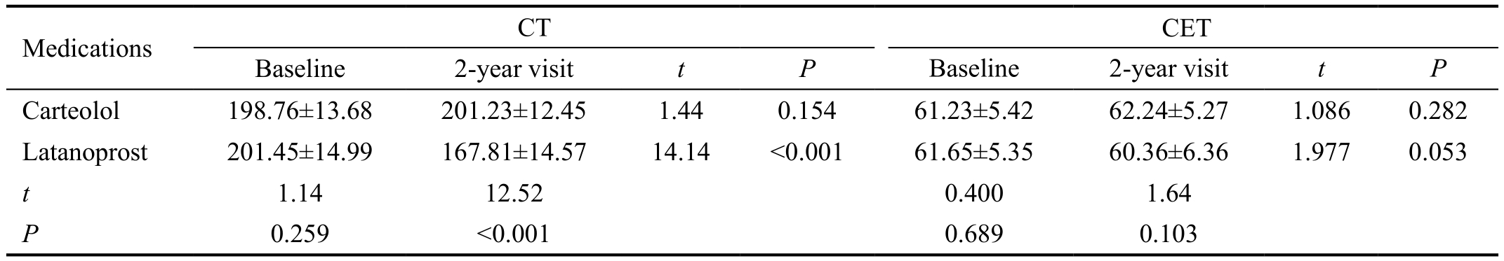
CT: Conjunctival thickness; CET: Conjunctival epithelial thickness; SD: Standard deviation.
CET Baseline 2-year visit t P Baseline 2-year visit t P Carteolol 198.76±13.68 201.23±12.45 1.44 0.154 61.23±5.42 62.24±5.27 1.086 0.282 Latanoprost 201.45±14.99 167.81±14.57 14.14 <0.001 61.65±5.35 60.36±6.36 1.977 0.053 t 1.14 12.52 0.400 1.64 P 0.259 <0.001 0.689 0.103 Medications CT
TreatmentOf the 106 eyes with no pre-administration of other anti-glaucoma eye drops, 55 eyes were treated with latanoprost eye drops (50 mg/mL, Xalatan; P fizer Manufacturing Belgium NV, Belgium) once a day (latanoprost group), while 51 eyes were treated with carteolol hydrochloride eye drops (2%,Carteolol, Otsuka Pharmaceutical Co., Ltd., China) twice a day(control group). All the included patients completed a followup of two years.
InstrumentationA Cirrus HD-OCT 4000 system (Carl Zeiss Meditec Inc., Dublin, CA, USA) was used in this study and cross-sectional bulbar conjunctival images were acquired with the Anterior Segment 5 Line Raster scanning protocol aimed at viewing high-resolution images of the anterior chamber angle and cornea.
Conjunctival Thickness and Conjunctival Epithelia Thickness MeasurementsThe method and procedures for CT measurement were previously reported in our studies[11].Briefly, subjects were positioned with their chin on the chin rest and forehead against the headrest with the scanned eye directed in an upper nasal gaze (left eye) or in an upper temporal gaze (right eye). This allowed the lower temporal conjunctiva 3-5 mm from the corneal limbus to be imaged.Using the clock analogy, the conjunctiva was imaged at 4:30 or 7:30. The Anterior Segment 5 Line Raster scan lines were then rotated to image the cross-sectional conjunctiva perpendicular to the limbus. When the images are digitally magnified, tissue landmarks can be identified by differences in brightness between tissues whereby the conjunctival epithelia thickness(CET) and CT can be measured (Figure 1). CT was measured using the central corneal thickness measurement software on the Cirrus OCT 4000. Five independent measurements of the temporal conjunctiva, each of which was the mean value of three measurements at three points separated by equal distance on the OCT image, were recorded and the mean value was used for statistical analysis.
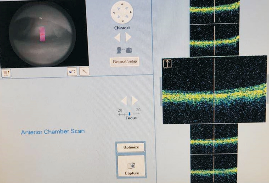
Fiugre 1 Conjunctival thickness was measured by OCT on a representative patient.
Both CET and CT were measured in all the recruited patients at presentation and at the designed endpoint (2y after first administration). All the thickness measurements were performed by two independent and masked observers.
Statistical AnalysisAll statistical analyses were performed using Stata 10.0 software (Stata Corp., College Station,TX, USA). A Shapiro-Wilk test was used to confirm normal distribution. The t-tests were then performed to determine whether there were significant differences in CT/CET between baseline and endpoint. A significance level of less than 0.05 was considered to significant.
RESULTS
The mean±standard deviation (SD) age in the latanoprost group was 68.84±7.84y, while it was 67.31±8.20y in the carteolol group. There were no difference in age between the two groups(t=0.9820, P=0.3284). At presentation, there was no difference in CET (t=0.400, P=0.689) or CT (t=1.14, P=0.259) between the two groups (Table 1). No significant difference was found in CET (61.65±5.35 μm at baseline, 60.36±6.36 μm at 2-year follow up, respectively; t=1.977, P=0.0531), while there was a significant decrease in CT from 201.45±14.99 μm at baseline to 167.81±14.57 μm at 2-year visit (t=14.1407, P<0.001) in the latanoprost group (Table 1; Figures 2, 3).
At 2-year follow-up, no statistically difference was found in CET (62.24±5.27 μm; t=1.086, P=0.282) or CT (201.23±12.45 μm;t=1.44, P=0.154) compared to it at baseline (CET: 61.23±5.42 μm;CT: 198.76±13.68 μm, respectively) in the carteolol group.(Table 1, Figures 4, 5).
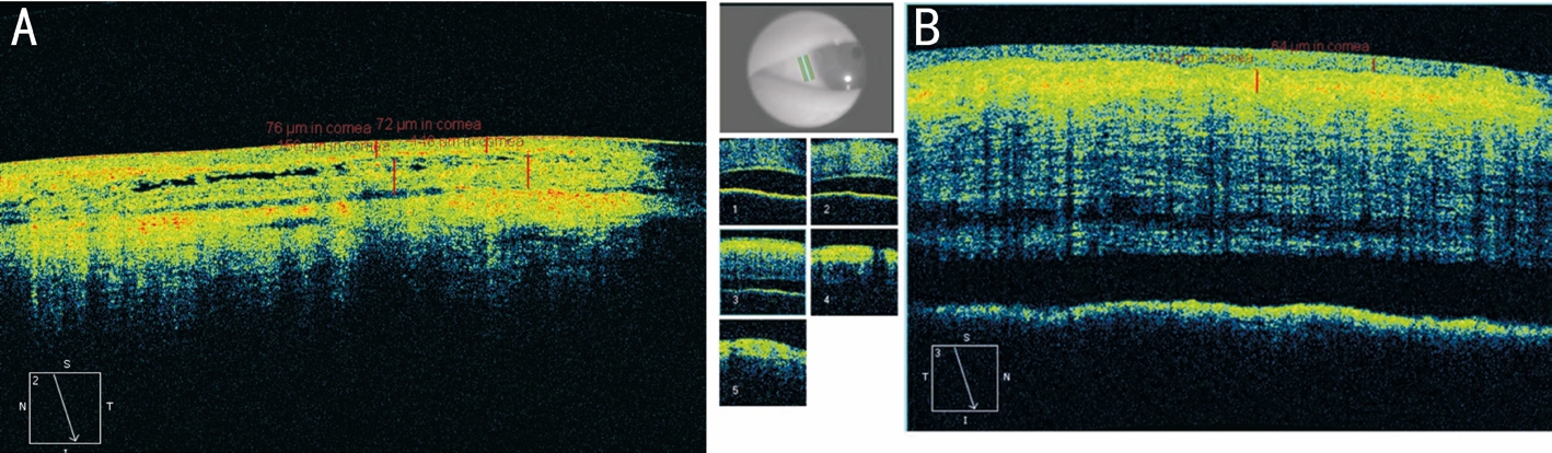
Figure 2 The representative OCT images showing CT and CET at baseline (A) and at 2-year follow up (B) in the latanoprost group.
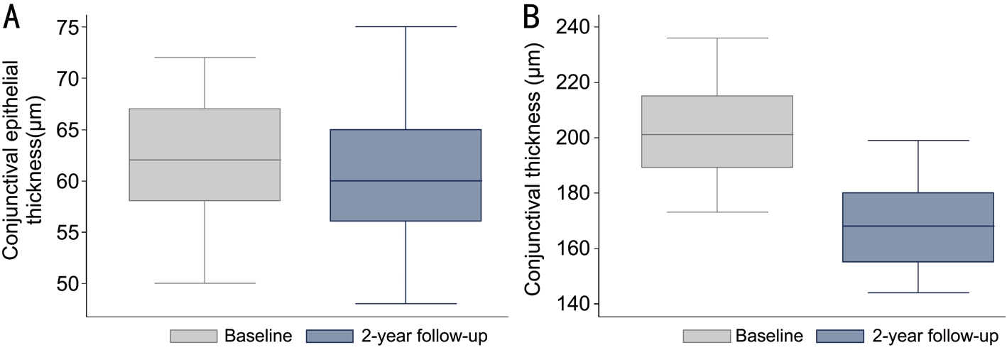
Figure 3 The comparisons of CET (A) and CT (B) between baseline and 2-year follow up in the latanoprost group.
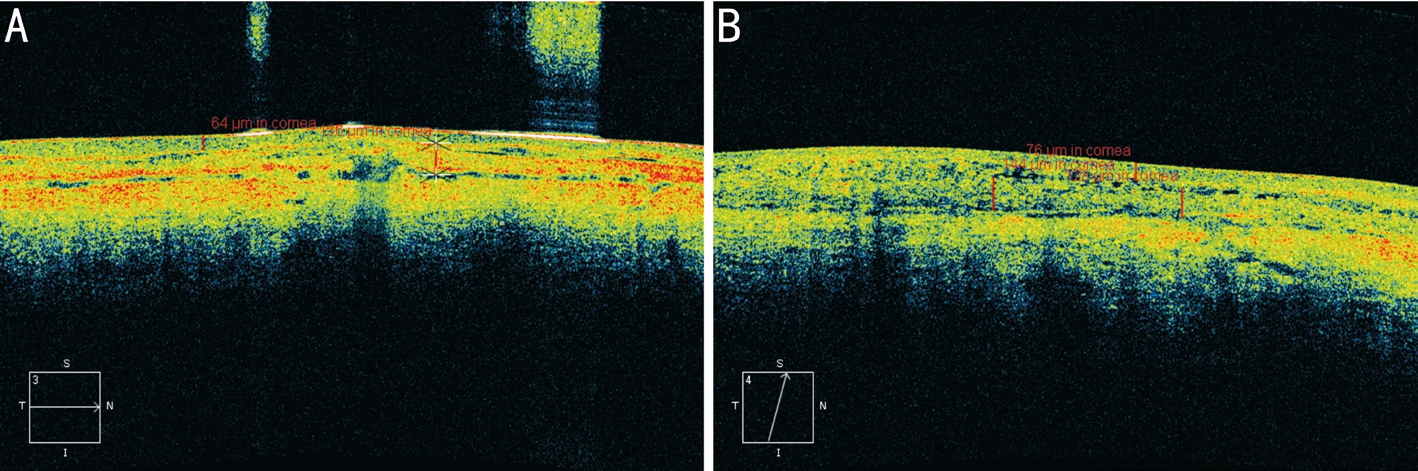
Figure 4 The representative OCT images showing CT and CET at baseline (A) and at 2-year follow up (B) in the carteolol group.
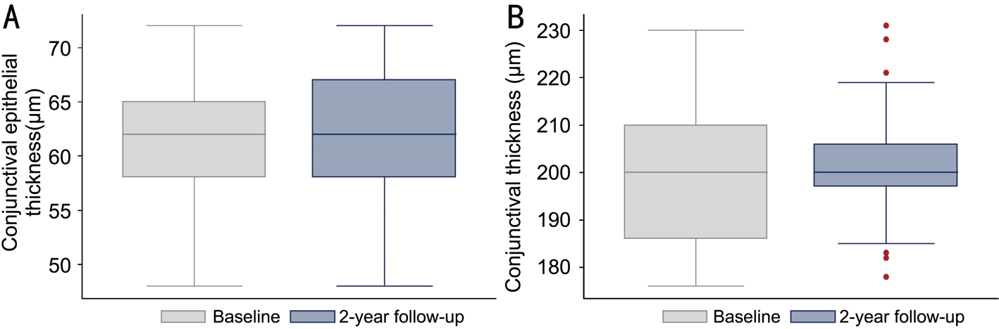
Figure 5 The comparisons of CET (A) and CT (B) between baseline and 2-year follow up in the carteolol group.
DISCUSSION
MMPs are a group of proteolytic enzymes responsible for catalysing extracelluar matrix degradation, the levels of which have been found in the ciliary body[12]. An experimental study in the latanoprost-treated specimens of monkey eyes has reported an upregulation of MMP-1 and MMP-2 and a decrease in collagen type IV and VI in the ciliary body[13],suggesting that latanoprost-induced increased activity of the extracellular matrix degradation might augment the flow of aqueous humor through the ciliary muscle bundles of the uveoscleral pathway and in turn lower IOP. The fact that MMPs have also been found in the conjunctiva[14]may argue for a mimicking change in the levels of MMPs in conjunctiva with topical prostaglandin analogs administration,as evidenced in a previous study in which an upregulation of MMP-1 and MMP-3 and a decreased collagen density were observed in the latanoprost-treated eyes[15]. A significant reduction of CT in the long-term latanoprost-treated glaucoma patients observed in our study may be a certain result of the up-regulated MMPs. Interestingly, in latanoprost-treated eyes,an increase of MMP-3 activity that may be a direct effect of latanoprost was found in the conjunctival epithelium[15], which seems to be inconsistent with the finding in our study in which no significant change in CET occurred to the eyes treated with topical latanoprost medication. The underlying reason for this disparity may be because of the lack of collagen fibers in the conjunctival epithelium.
Incisional surgery to lower IOP is still indicated for those who fail medicine treatments. However, scarring and extracelluar matrix accumulation within the subconjunctival space may confer treatment failure, for which, the stimulation of extracelluar matrix degradation that suppresses subconjunctival scar formation and promote longer survival of filtering blebs may have a positive effect[16]. In this context, the accompanying effect of conjunctival thinning in the wake of long-term latanoprost medication seems to be helpful in maintaining the function of filtering blebs. However, the outcome of filtration surgery is determined as much by the performance of filtration bleb as by how the drainage pathways in conjunctiva would perform to drain aqueous humor from subconjunctival space after surgery. The conjunctival lymphatics, among others, have been confirmed to play an important role in determining the surgical outcome in glaucoma filtration surgery in rabbits and monkeys[17]. A previous study has demonstrated that bulbar conjunctiva is rich in lymphatics that consist of abundant initial lymphatics, fewer communication branches, and some precollectors; the majority of the initial lymphatics are located in the super ficial conjunctiva between the epithelium and Tenon’s capsule[18]. Despite there being no knowledge available,it is logical that a reduction in the density of conjunctival lmyphatics may occur with the thinning of conjunctiva. With this understanding, long-term topical latanoprost medications may have detrimental effect on the filtration surgery. Although latanoprost has assumed a large role in lowering IOP for glaucoma therapy, its potential effect on the filtration surgery is not fully understood and calls for further studies to investigate.An increase of tissue inhibitor of matrix metalloproteinases(TIMPs), the endogenous inhibitors of MMPs, was found in the aqueous humor of POAG-affected eyes and it is concluded that the increased collagen synthesis by the upregulation of TIMPs may contribute to an increased deposition of collagen in the trabecular meshwork and thus play a role in the pathogenesis of POAG[19]. If the same is true of the conjunctiva and the already existing high levels of TIMPs at presentation may have attenuated the true effect of latanoprost-induced CT thinning; in this study, a group of patients receiving the treatment of carteolol were recruited as the control group in which no significant change in CET or CT was found across a 2-year follow-up-something that nevertheless cannot answer this question. To this end, a study aimed at investigating CT in POAG patients and in normal individuals may provide some information on this issue. However, this is not the topic of the present study.
In conclusion, the first in vivo study has presented a significant decrease in CT in glaucoma patients treated with long-term topical latanoprost; its potential effect on the outcome of filtration surgery should be considered.
ACKNOWLEDGEMENTS
Foundations:Supported by a grant from Putuo Hospital affiliated to Shanghai University of Traditional Chinese Medicine (No.2071301A); a fund program from Shanghai University of Traditional Chinese Medicine for incubation of doctor degree (No.B 201708/J-16-11).
Conflicts of Interest: Li QS,None;Bao FF,None;Zhang ZY,None;Ma K,None.
REFERENCES
1 O’Callaghan J, Cassidy PS, Humphries P. Open-angle glaucoma:therapeutically targeting the extracellular matrix of the conventional out flow pathway.Expert Opin Ther Targets2017;21(11):1037-1050.
2 Zhong Y, Gao J, Ye W, Huang P, Cheng Y, Jiao Q. Effect of latanoprost acid and pilocarpine on cultured rabbit ciliary muscle cells.Ophthalmic Res2007;39(4):232-240.
3 Vicente A, Prud’homme S, Ferreira J, Abegão Pinto L, Stalmans I.Open-angle glaucoma: drug development pipeline during the last 20 years(1995-2015).Ophthalmic Res2017;57(4):201-207.
4 Schrems WA, Schrems-Hoesl LM, Mardin CY, Horn FK, Juenemann AG,Kruse FE, Braun JM, Laemmer R. The effect of long-term antiglaucomatous drug administration on central corneal thickness.J Glaucoma2016;25(3):274-280.
5 Yoo R, Choi YA, Cho BJ. Change in central corneal thickness after the discontinuation of latanoprost in normal tension glaucoma-change in central corneal thickness after stop of latanoprost.J Ocul Pharmacol Ther2017;33(1):57-61.
6 Sen E, Nalcacioglu P, Yazici A, Aksakal FN, Altinok A, Tuna T, Koklu G. Comparison of the effects of latanoprost and bimatoprost on central corneal thickness.J Glaucoma2008;17(5):398-402.
7 Arcieri ES, Pierre Filho PT, Wakamatsu TH, Costa VP. The effects of prostaglandin analogues on the blood aqueous barrier and corneal thickness of phakic patients with primary open-angle glaucoma and ocular hypertension.Eye (Lond)2008;22(2):179-183.
8 Hatanaka M, Vessani RM, Elias IR, Morita C, Susanna R Jr. The effect of prostaglandin analogs and prostamide on central corneal thickness.J Ocul Pharmacol Ther2009;25(1):51-53.
9 Terai N, Schlötzer-Schrehardt U, Lampel J, Böhm AG, Rummelt C, Schmidt E, Pillunat LE. Effect of latanoprost and timolol on the histopathology of the human conjunctiva.Br J Ophthalmol2009;93(2):219-224.
10 Zhang X, Li Q, Xiang M, Zou H, Liu B, Zhou H, Han Z, Fu Z, Zhang Z, Wang H. Bulbar conjunctival thickness measurements with optical coherence tomography in healthy Chinese subjects.Invest Ophthalmol Vis Sci2013;54(7):4705-4709.
11 Zhang XR, Zhang ZY, Hoffman MR, Li QS, Liu B, Zhou HM. The effect of age and conjunctivochalasis on conjunctival thickness.Curr Eye Res2013;38(3):331-334.
12 Oh DJ, Martin JL, Williams AJ, Peck RE, Pokorny C, Russell P,Birk DE, Rhee DJ. Analysis of expression of matrix metalloproteinases and tissue inhibitors of metalloproteinases in human ciliary body after latanoprost.Invest Ophthalmol Vis Sci2006;47(3):953-963.
13 Ocklind A. Effect of latanoprost on the extracellular matrix of the ciliary muscle. A study on cultured cells and tissue sections.Exp Eye Res1998;67(2):179-191.
14 Ito T, Ohguro H, Mamiya K, Ohguro I, Nakazawa M. Effects of antiglaucoma drops on MMP and TIMP balance in conjunctival and subconjunctival tissue.Invest Ophthalmol Vis Sci2006;47(3):823-830.
15 Mietz H, Schlötzer-Schrehardt U, Strassfeld C, Krieglstein GK. Effect of latanoprost and timolol on the histopathology of the rabbit conjunctiva.Invest Ophthalmol Vis Sci2001;42(3):679-687.
16 Addicks EM, Quigley HA, Green WR, Robin AL. Histologic characteristics of filtering blebs in glaucomatous eyes.Arch Ophthalmol1983;101(5):795-798.
17 Yu DY, Morgan WH, Sun X, Su EN, Cringle SJ, Yu PK, House P,Guo W, Yu X. The critical role of the conjunctiva in glaucoma filtration surgery.Prog Retin Eye Res2009;28(5):303-328.
18 Guo W, Zhu Y, Yu PK, Yu X, Sun X, Cringle SJ, Su EN, Yu DY.Quantitative study of the topographic distribution of conjunctival lymphatic vessels in the monkey.Exp Eye Res2012;94(1):90-97.
19 González-Avila G, Ginebra M, Hayakawa T, Vadillo-Ortega F, Terán L,Selman M. Collagen metabolism in human aqueous humor from primary open-angle glaucoma. Decreased degradation and increased biosynthesis play a role in its pathogenesis.Arch Ophthalmol1995;113(10):1319-1323.
Co- first authors:Qing-Song Li, Fang-Fang Bao and Kai Ma
Correspondence to:Zhen-Yong Zhang. No. 164, Lanxi Road,Shanghai 200060, China. zzyly818@sina.com
Received:2018-01-23 Accepted: 2018-04-23
Abstract ● AlM: To investigate the effect of long-term use of topically administered latanoprost on conjunctival thickness (CT) and conjunctival epithelium thickness (CET)in the patients with glaucoma.● METHODS: A series of 106 glaucomatous patients were included. Of the 106 eyes, 55 eyes were treated with latanoprost eye drops once a day (latanoprost group),while 51 eyes were treated with carteolol hydrochloride eye drops (carteolol group). All the included patients completed a 2-year follow-up. CT and CET were measured with optical coherence tomography (OCT) in all patients at presentation and at 2-year visit, respectively. Statistical analysis was then performed to compare the change in CT and CET.● RESULTS: At presentation, there was no difference in CET (t=0.400, P=0.689) or CT (t=1.14, P=0.259) between the two groups. No significant difference was found in CET (61.65±5.35 μm at baseline, 60.36±6.36 μm at 2-year follow-up, respectively; t=1.977, P=0.0531), while there was a significant decrease in CT from 201.45±14.99 μm at baseline to 167.81±14.57 μm at 2-year visit (t=14.1407,P<0.001) in the latanoprost group. At 2-year follow-up, no statistically difference was found in CET (62.24±5.27 μm;t=1.086, P=0.282) or CT (201.23±12.45 μm; t=1.44, P=0.154)compared to it at baseline (CET: 61.23±5.42 μm; CT:198.76±13.68 μm, respectively) in the carteolol group.● CONCLUSlON: A significant decrease in conjunctival thickness is found in glaucoma patients treated with long-term topical latanoprost; its potential effect on the outcome of filtration surgery should be considered.
● KEYWORDS:conjunctival thickness; latanoprost; glaucoma;optical coherence tomography; glaucoma surgery
DOl:10.18240/ijo.2018.07.14
Citation:Li QS, Bao FF, Zhang ZY, Ma K. Effect of long-term topical latanoprost medication on conjunctival thickness in patients with glaucoma. Int J Ophthalmol 2018;11(7):1158-1162