
・Letter to the Editor・
A
tooth lesion in the eyelid in a Chinese infant: a rare case report
Jian Liu, Jun Chen, You-Sheng Zhang, Jie Zhu, Xiao Lin,
Yuan-Jian Wei
Department
of Ophthalmology, Affiliated Nanping First Hospital of Fujian Medical
University, Nanping 353000, Fujian Province, China
Correspondence
to: Jun Chen.
Department of Ophthalmology, Affiliated Nanping First Hospital of Fujian
Medical University, 317 zhongshan Road, Nanping 353000, Fujian Province, China.
m18776769056@163.com
Received:
DOI:10.18240/ijo.2019.11.22
Citation:
Liu J, Chen J, Zhang YS, Zhu J, Lin X, Wei YJ. A tooth lesion in the eyelid in
a Chinese infant: a rare case report. Int J Ophthalmol
2019;12(11):1807-1808
Dear Editor,
My name is Jun Chen and I am
currently working as an ophthalmologist at the Affiliated Nanping First
Hospital of Fujian Medical University. I am writing this letter to present a
case of a tooth lesion in the eyelid.
Choristomas are believed to be
developmental malformations of normal, mature-appearing tissues in an abnormal
anatomic location[1]. Choristomas consist of
dermis-like tissue or ectopic tissues of mesoectodermal origin (lacrimal and
other glands, fat, nerve, brain, cartilage, bone, and teeth)[2].
An ectopic tooth in the eyelid was first reported by Van Der Straeten[3] in 1934, then reported by Subramaniam et al[4] in 1966 and finally by Jakobiec et al[5] who named it palpebral odontogenic choristoma in 2009.
Here, we describe a case of a tooth lesion in the eyelid in a Chinese infant.
According to Declaration of Helsinki, a written voluntary informed consent was
obtained from the patient for publication of this case report and accompanying
images.
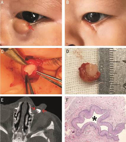
In March
2018, a 7-month-old baby girl was referred to our hospital for further
management of congenital tumor and malformation in the lower inner eyelid of
the left eye. The girl was a premature infant. There was no family history of
ocular diseases or ocular trauma.
On
examination at our clinic, slit-lamp examination showed a 15×
Figure
With a
suspected clinical diagnosis of congenital tumor and malformation, the mass was
removed totally under general anesthesia and sent for pathologic examination in
March 23, 2018 (Figure 1B, 1D). During this surgery, a full-thickness resection
of the eyelid was undertaken. Due to infringement of mass, the interior punctum
and canaliculus was undertaken too. Palpebral dissection revealed a tooth
(Figure
An ectopic
tooth in the eyelid is extraordinarily rare. Since it was first reported by the
French ophthalmologist Van Der Straeten[3] in
1934, only few cases have been previously reported in the literature[3-5]. To our knowledge, this is the
fourth case report of a tooth structure discovered in the eyelid and not been
previously reported in a Chinese population.
The previous
three cases plus the present one all occurred in the inner aspect of the lower
eyelid. Moreover, all 4 cases have described as dome-shaped, globoid eyelid
eminences with adjacent eyelid thickening. In the previous cases, the mass was
firm but it was soft in our case. The reason may be that ectopic tooth was
located in underlying tissue in our case. As the case reported by Jakobiec et
al[5], the mass also involved the eyelid
margin and eyelashes were lost in our case. The most closely mimicking entity
was the phakomatous choristoma which should be considered as the first
differential diagnosis[5].
Choristomas
are occasionally familial[2]. But in this case, we
did not have any meaningful findings.
In August
2018, we tried to obtain a panoramic dental X-ray at visit. But it was failed
because the infant could not cooperate.
Due to our
mistakes, when pathologist processes the mass, the tooth was discarded
regrettably, and the surrounding soft tissue was collected only. As a result,
there are less histopathological details and photomicrographs in this case.
In conclusion, this is the first
described case of an ectopic teeth within the eyelid in a Chinese patient in
the literature. In this case, globoid congenital tumor in the lower inner
eyelid of the left eye and a punctum-like structure is observed, CT showed that
abnormal high-density shadow with calcification near the lacrimal sac. Under
general anesthesia, the mass was removed totally.
ACKNOWLEDGEMENTS
Conflicts of
Interest: Liu J,
None; Chen J, None; Zhang YS, None; Zhu J, None; Lin X,
None; Wei YJ, None.
REFERENCES