bannter.jpg
·
Review Article·


Combined corneal CXL and photorefractive keratectomy for treatment of keratoconus: a review


Mansour M. Al-Mohaimeed


Department of Ophthalmology, College of Medicine, Qassim University, Qassim, Buraidah 51452, Kingdom of Saudi Arabia

Correspondence to: Mansour M. Al-Mohaimeed. Department of Ophthalmology, College of Medicine, Qassim University, Qassim, PO Box 6655, Buraidah 51452, Kingdom of Saudi Arabia. drmohaimeed@qumed.edu.sa

Received: 2019-03-01        Accepted: 2019-05-29


Abstract

Keratoconus and iatrogenic keratectasia are the corneal ectatic disorders occurring due to biomechanical weakening of the cornea resulting in distorted images, myopia, and irregular astigmatism. Corneal collagen cross-linking (CXL) is performed to arrest keratoconus successfully. The main aim of this review is to discuss the safety and efficacy of the adjuvant therapies, such as the combination of CXL and photorefractive keratectomy (PRK) for the treatment of corneal ectatic disorders. A comprehensive literature search was performed using PubMed, MEDLINE, and Scopus using keywords ‘collagen’ ‘keratoconus’, ‘keratectasia’, ‘collagen cross-linking’, and ‘photorefractive keratectomy’. Search results were restricted to clinical studies published in English. Corneal CXL effectively arrests the progression of keratoconus by enhancing corneal rigidity. However, functional vision is not improved by cross-linking. Combining CXL to refractive surgeries such as topography-guided PRK or transepithelial PRK is found to be a safe and effective method in providing corneal stability as well as significantly improving functional visual acuity with few minor complications. This combined technique also prevents regression of keratoconus and reduce the risk of keratectasia. CXL combined with PRK is a promising therapeutic approach in ophthalmology that can be successfully used to treat progressive keratoconus and other corneal ectatic disorders and to enhance visual acuity.

KEYWORDS: corneal collagen cross-linking; photorefractive keratectomy; keratoconus

DOI:10.18240/ijo.2019.12.16


Citation: Al-Mohaimeed MM. Combined corneal CXL and photorefractive keratectomy for treatment of keratoconus: a review. Int J Ophthalmol 2019;12(12):1929-1938


INTRODUCTION

Keratoconus is a bilateral, non-inflammatory, progressive ectatic disease characterized by apical bulging of the cornea, thinning of the central cornea, and distortion of the cornea, which affects mostly adolescent people[1-2]. With the advancement of the disease, ocular aberrations increase, and image quality and visual acuity are diminished. In severe cases, axial corneal scarring and irregular astigmatism were also noticed. The key objective of the treatment of keratoconus involves halting the progression of ectasia, improving the refractive errors, and bringing back the normal shape of the cornea[3]. Progressive keratectasia resulting from corneal disease or a sequela of laser in situ keratomileusis (LASIK) surgery has no appropriate treatment at present. Available treatment of keratoconus mostly involves interventions done for tectonic, optical, or refractive purposes. Treatment of keratoconus depends on the extent of disease progression and disease severity[3]. Conventional approaches to treating mild to moderate keratoconus involve eyeglasses and rigid gas permeable contact lenses[3]. Nonetheless, some patients are unable to tolerate contact lens and spectacle correction is insufficient in some cases. Moreover, in advanced stages of keratoconus with excessive corneal thinning/steepening and corneal scarring[4], the traditional treatment approaches are not quite effective. Furthermore, none of these therapeutic approaches are able to treat the principal causes of ectasia and do not guarantee the absolute cessation of keratoconus progression[5]. One promising treatment approach gaining popularity from the late decades of the twentieth century is the corneal collagen cross-linking (CXL), which aimed to treat the underlying pathology of keratoconic eyes and effectively stiffens the cornea by restoring its tensile strength[4] and subsequently slow down or arrest the advancement of keratoconus, or even reverses keratoconus in rare cases[5-7]. Additionally, a combination of CXL with photorefractive keratectomy (PRK), a standard laser-assisted refractive surgery is expected to have greater efficacy in the management of keratoconus. The main purpose of the combined treatment of keratoconus with PRK/CXL involves strengthening the cornea and halting the disease progression by CXL and to improve the quality of vision via PRK[4].

The current paper intends to review recent literature on the application of corneal CXL in combination with PRK for treating keratoconus and other corneal ectatic disorders.

Basic Principles of CXL  Collagen is a triple helical structural protein present abundantly in the extracellular matrix in all animals. Intermolecular cross-links between collagen monomers aid in strengthening the collagen structure. CXL is a natural phenomenon occurring within the corneas and crystalline lens either enzymatically or non-enzymatically. The enzymatic cross-linking occurs via lysyl oxidase enzyme[8]. Non-enzymatic cross-linking occurs via glycation, where bond formation occurs between sugar and the amino group of a protein; this mechanism commonly occurs with age, or in an individual with diabetes mellitus, thereby strengthens the cornea in elderly people and lowers the occurrence of keratoconus in diabetes mellitus patients. CXL can also be induced by oxidation using ultraviolet (UV) irradiation to generate reactive oxygen species (ROS) that polymerize the collagen monomers into cross-linked polymers. The effect of CXL reduces with low oxygen tension indicating the importance of oxygen and ROS in collagen polymerization[4].

History of CXL  The most common application of CXL is to fix tissue and strengthening the heart valve. CXL emerged from researches conducted to detect biological glues to make cornea strong. The scientists intended to obtain corneal cross-linking in non-diabetic corneas analogous to natural cross-linking by glycosylation in diabetic patients[7]. Finally, in 2003, Wollensak et al[9] introduced the CXL technique using 370 nm UVA irradiation and photomediator riboflavin to cross-link stromal collagen fibrils for treating keratoconus[7]. This technique is widely followed at present. Food & Drug Administration (FDA) in the USA also approved the use of CXL in 2016 for treating progressive keratoconus and the post-LASIK ectasia based on the results of three 12-month clinical trials[4].

Use of Riboflavin in Corneal CXL with UVA  Corneal CXL is a minimally invasive method of cross-linking corneal collagen in order to enhance the biomechanical stability of the cornea, which is weakened due to progressive keratoconus or post-operative keratectasia[10]. In this method, riboflavin or vitamin B2 (a photosensitizing substance) and UVA are used to form additional intra and inter-fibrillar covalent bonds via photosensitized oxidation[11]. Riboflavin treated corneas have three absorption peaks- 270, 365, and 370 nm. The peak between 365 and 370 nm is normally used in the CXL procedure as this does not damage the retina[12]. Riboflavin is excited into a triplet state by UVA light of wavelength 370 nm[12] and produces ROS to activate natural lysyl oxidase pathway[5]. The increased cross-links between and within collagen fibers stabilize the stromal collagen fibers, thereby improving the collagen structure and corneal rigidity[10] and resist it from deformation[13]. The use of 0.1% riboflavin in CXL technique has been found to enhance corneal UVA absorption by 95% compared to 30% when UVA was used alone. Moreover, riboflavin reduces keratocyte cytotoxicity caused by UVA[8,10]. Furthermore, riboflavin is anticipated to serve as a protective layer of the cornea, which may even reach up to 400 µm following 30min application, and protect the internal structures such as the retina, crystalline lens, and the endothelium from the harmful effects of UVA[11].

Techniques of Corneal CXL  Wollensak and Spoerl first developed a photochemical CXL procedure at the University of Dresden, commonly referred to as the Dresden protocol[9]. Till date many protocols have been recommended for corneal cross-linking; however, the basis of all these is the Dresden protocol established by Wollensak et al[9]. The entire procedure is conducted under sterile condition. Corneal CXL begins with the removal of corneal epithelium since the epithelial tight junctions block riboflavin absorption to some extent. De-epithelization results in uniform riboflavin diffusion in the corneal stroma[6]. Under topical anesthesia, abrasion of the central 7-9 mm of the corneal epithelium is performed followed by administration of 0.1% riboflavin solution in 10 mL of 20% dextran and 10 mg riboflavin-5-phosphate in the cornea for about 30min to permeate cornea before UVA irradiation[6] (Figure 1). Riboflavin, being a photomediator enhances corneal UVA absorption. An optical system providing uniform beam of UVA irradiation is essential for CXL to allow proper UVA absorption by riboflavin and thereby causing effective cross-linking[4,12]. UVA light of wavelength 370 nm and irradiance of 3 mW/cm2 is applied for 30min at a distance of 5.4 mm from the cornea, thus delivering a dose of 5.4 J/cm2[4,6]. Riboflavin solution and balanced salt solution are injected during UVA irradiation to saturate and hydrate the cornea. Once the treatment is completed, antibiotic eye drop is applied and a bandage contact lens is placed till complete reepithelization[11].

Mansour M

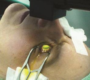
Figure 1 Treatment in progress with the cornea soaked with riboflavin and irradiated by the UV lamp (represented from Jankov et al[11]).


Clinical Study Results with Standard CXL Procedure  Wollensak[14] performed the first clinical study of corneal CXL in 2003. This 3-year study detected that following CXL treatment in patients with advanced keratoconus, the progression of keratoconus was stopped in all patients along with improvement in best corrected visual acuity (BCVA). Since then, a multitude of clinical studies including prospective as well as retrospective studies has been performed to explore the effectiveness of the standard CXL procedure. The main parameters evaluated at the follow-up treatment are the maximal keratometry (Kmax) value, BCVA, uncorrected distance visual acuity (UDVA) and the follow-up period usually ranged between 1 and 6y[15]. Raiskup and colleagues in a retrospective study determined the long-term efficacy of CXL in the stabilization of keratoconus with a significant reduction of Kmax and Kmin values and also improvement in BCVA. Another study with the largest follow-up time (48mo), although detected initial deterioration (first 6mo), later found a substantial improvement in next 42mo[4]. The results of the majority of the clinical studies revealed that standard CXL has stabilized corneal keratometry and improved BCVA, UDVA, visual acuity, and topographical indexes in keratoconic eyes without altering corneal volume and anterior chamber volume and depth[16]. Some studies reported about improvement of visual acuity but no change in keratometry values, whereas few other studies stated minor reduction in UDVA and BCVA readings after 4-5y of CXL treatment. In majority of the cases, diminution of irregular astigmatism was responsible for better visual acuity[16].

There are not much randomized controlled trials to clarify the results of these studies. Nevertheless, the findings of the first randomized clinical trial on the use of CXL in treating progressive keratoconus conducted by Witting-Silva et al[17] with a follow-up period of three years substantiated the effectiveness of standard CXL protocol in stabilizing keratoconus progression and is considered to be a notable landmark. Another prospective, non randomized clinical study on CXL for treating progressive keratoconus determined statistical improvement in visual acuity and statistically significant reduction of Kmax and Kmin values in the treated group versus untreated group with no major change in endothelial cell count at 12-month follow-up[12]. The long-term results (48-60mo follow-up) of an open, prospective, nonrandomized, Phase II clinical trial conducted by Caporossi et al[18] also determined stability or improvement in 92% cases with a mean reduction of average keratometry readings and substantial improvement in visual acuity, BCVA, and UCVA following standard CXL, whereas the untreated fellow eyes showed 65% progression of keratoconus within 2y.

Treatment Failure  Treatment failure is defined as the continual progression of keratoconus with an enhancement of Kmax reading of 1.0 D over the preoperative value. Treatment failure has been found to occur in 8.1%-33.3% cases; one study by Poli et al[19] stated about 11% failure rate during a follow-up period of 6y.

Complications of Standard CXL  Corneal CXL is a relatively safe and effective revolutionary therapeutic approach to pause keratoconus progression for at least five years and postoperative LASIK ectasia for a minimum of two years with a low rate of complications[20]. The complications of CXL can be either primary or direct arising from an incorrect application of the technique or incorrect patient selection. The secondary or indirect complications of CXL result from patient’s poor hygiene, therapeutic soft contact lens, or other ocular surface diseases, such as bacterial keratitis occurring due to epithelial defect or use of soft bandage contact lens following surgery. The two most common direct complications of CXL include 1) appearance of stromal haze due to back-scattered and reflected light; 2) corneal edema due to endothelial damage[6]. Previous studies reported that CXL-associated corneal haze appearing as a dust-like change in corneal stroma actually differs from other types of corneal haze; this postoperative corneal haze usually increases within 1-3mo of surgery and by 6mo, haze diminishes and the cornea appears to be clear[21].

Typically, corneal endothelial damage occurs when safety limits about corneal thickness are not followed. CXL results in corneal thinning, which starts at the initial phase of the procedure and continues until 1-3mo’ post-treatment. Nonetheless, the optimal healing and remodeling of the cornea occur in the first 6mo to 1-year period. In fact, corneal thickness begins to recover from 3mo and attains baseline thickness (i.e. corneal thickness before CXL procedure) within 1y. However, Kim et al[22] in their study reported statistically significant reduction in the corneal thickness as compared to baseline value even 5y post-CXL treatment. Corneal thinning following CXL probably occurs due to corneal desiccation and dehydration owing to prolonged UVA exposure and this actually results in endothelial damage[16]. The endothelial damage can be prevented by keeping the corneal thickness over 400 µm prior to UV exposure[21].

Modifications of Conventional CXL Technique  Conventional CXL technique is contraindicated for individuals with corneas thinner than 400 μm[5] in order to protect the cornea from endothelial toxicity and cell death[23]. Hence, CXL using standard protocol is proposed for keratoconic eyes with corneal thickness at least 400 μm following de-epithelization. Progression has been reported in about 25%-30% of keratoconus cases[23]. In order to overcome the possible complications arising from the use of standard CXL technique in keratoconus patients who are not good candidates for traditional CXL (eyes with corneal thickness less than 400 μm) or to obtain quicker results, several modifications have been made in the conventional Dresden protocol[24]. The common modifications include: 1) use of hypoosmolar riboflavin to swell thin corneas artificially; 2) accelerated CXL, altering irradiation dosage to reduce treatment duration; and 3) transepithelial CXL (TE-CXL), keeping epithelium intact and using various compounds to enhance riboflavin penetration[25].

CXL with hypoosmolar riboflavin  Original Dresden protocol mentions the use of 0.1% riboflavin in 20% dextran solution. This riboflavin concentration can treat only anterior 300 μm of the stroma and is ineffective when corneal pachymetry is <400 μm after de-epithelization. A permanent stromal scar was noticed in keratoconic eyes with thinner corneas and steeper keratometric values following CXL using isomolar riboflavin[23]. In contrast to isotonic riboflavin, hypoosmolar riboflavin has lower colloidal pressure (402.7 mOsmol/L vs 310 mOsmol/L) that causes stromal swelling to double its thickness where stromal bed is less than 400 μm and thus facilitates CXL technique[23,26]. In a study, Wollensak et al[9] used hypoosmolar riboflavin alone in every 2min for 30min in kertaoconic eyes with thin corneas (<400 μm) and observed stability in vision and keratometry with no stromal scars at 12mo’ follow-up. Hafezi et al[10] performed CXL in progressive keratoconus patients (cornea <400 μm) using hypoosmolar riboflavin and detected halting of keratoconus progression in all patients along with stable keratometry at 6-month follow-up. Stojanovic et al[27] noticed that use of hypoosmolar riboflavin with standard irradiation of 3 mW/cm2

for 30min arrested keratoconus progression; however, the efficacy was lower than traditional CXL with isotonic riboflavin. The possible explanation is that in hydrated corneas (using hypoosmolar robiflavin) concentration of collagen fibrils is diminished, hence fewer collagen fibrils are available for CXL[23].

Accelerated versus conventional CXL in treating keratoconus  The duration of standard CXL is about 1h and exposure of the cornea to UVA for this time period may cause damage to corneas thinner than 400 μm. To quicken the treatment process, “accelerated CXL” is performed. This technique utilizes high energy up to 30 mW/cm2 for a shorter duration of time such as 3-10min, still keeping the total radiant exposure to be 5.4 J/cm2[2]. Several studies were conducted to compare the efficacy of accelerated CXL with that of conventional CXL by using different irradiation intensity and it was observed that Accelerated protocols have acceptable efficacy[4]. However, a recent study comparing accelerated vs conventional CXL in keratoconic eyes was unable to detect any significant difference in visual acuity, keratometry reading, and endothelial cell count at 1-year follow-up among these two techniques[26].

Transepithelial CXL vs conventional epithelium-off CXL  Wollensak et al[9] performed CXL by excision of corneal epithelium to facilitate penetration of riboflavin since riboflavin being hydrophilic unable to penetrate properly through the lipophilic epithelial membrane. However, removal of epithelium is a painful method, requires more healing time, has a higher probability of developing infections, and leads to corneal melting[4]. To minimize these problems, currently, a modified CXL technique known as TE-CXL, where corneal epithelium remains intact is being performed[4]. The entry of riboflavin through corneal epithelium is aided by the addition of certain chemicals such as tetracaine, benzalkonium chloride, and trometamol, which loosen the epithelial tight junctions[4,11]. Stojanovic et al[27] did a comparative study with and without epithelial removal to treat progressive keratoconus and concluded that both methods were equally safe and effective in stabilization of keratoconus. While different studies revealed that visual acuity appears to be similar following TE-CXL and epithelium-off CXL, the efficacy of TE-CXL in terms of topographic indices is less than CXL with de-epithelization[4,11]. In one study limited CXL effect was observed in eyes with intact epithelium; the possible reasons may be insufficient riboflavin concentration in the stroma and lesser oxygen diffusion into the stroma. It is anticipated that rise in biomechanical rigidity following TE-CXL and standard epithelium-off CXL is about 64% and 320% respectively[23] suggesting that the effect of TE-CXL is more superficial than conventional CXL[26].

Despite this, TE-CXL has several advantages over regular epithelium-off CXL, including less time-consuming, no operation room required, quicker visual recovery, applicable for patients with corneal thickness less than 400 μm, safer technique since intact cornea acts as a barrier to prevent the entry of pathogen, reducing the occurrence of infectious keratitis[4]. In addition, stromal haze, postoperative pain, burning sensation, healing reaction, and other complications are less in TE-CXL[23,27].

Iantophoresis-assisted CXL  Riboflavin is a crucial component of CXL since by virtue of its photosensitizing power, it forms the CXL and provides tensile strength to cornea. Thus, proper penetration of riboflavin to the stroma is vital. Iantophoresis is a non-invasive unique technique to facilitate riboflavin infiltration using small electric current. Riboflavin, being negatively charged is a good candidate for iontophoresis. Following only 5min of 1 mA current flow, an adequate level of riboflavin penetrates into the corneal stroma, thus epithelial integrity is maintained[15,23]. Initial clinical study results exhibited that iontophoresis-assisted CXL can stop keratoconus advancement without considerable complications; even so, further long-term follow-up studies are needed to determine its efficacy in keratoconus management[15].


PHOTOREFRACTIVE KERATECTOMY IN COMBINATION WITH CXL TO TREAT KERATOCONUS AND POST-LASIK KERATECTASIA

Corneal CXL is a promising technique for management of keratoconus as it provides tensile strength and stability to the cornea by inducing cross-links at the corneal stroma and thus arrests keratoconus. For prophylactic use, virtually any patient can be treated with cross-linking to reduce the chance of future development of ectasia, especially patients with thinner than normal corneas, irregular corneal astigmatism, asymmetry on corneal topography, against-the-rule astigmatism or steeper than normal corneas. Majority of the studies indicated more than 90% success rate in stabilizing the advancement of keratoconus following CXL technique[28]. However, CXL alone is unable to improve functional vision[29] and yields a better result for the patients suffering from early-to-moderate keratoconus compared to end-stage keratoconus[6]. The limitation of CXL can be resolved by combining CXL with PRK. Although previous studies revealed the effectiveness of PRK in treating stable or early keratoconus, its application in combination with CXL proved to be superior[30]. Presently, a novel technique referred to as ‘CXL Plus’, which combines two surgical procedures is performed to treat a corneal ectatic disorder such as keratoconus. In CXL-Plus, the fundamental method is CXL, which is combined with other refractive procedures such as topography-guided PRK, or transepithelial topography-guided PRK, or intracorneal ring segments (ICRS), or phakic intraocular lens implantation (PIOL), or multiple techniques combined with CXL, either sequentially or simultaneously[29,31]. CXL-Plus is advantageous over typical CXL because it enhances CXL result, by improving corneal stability as well as by providing functional visual acuity[29]. Topography-guided PRK combined with CXL was the first CXL Plus method using excimer laser ablation and is considered to be an effective treatment of choice for keratoconus and keratectasia[29]. Several modifications of the technique have been suggested, involving the timing of the two procedures (simultaneous or sequential), highest advised ablation depth, and the use of mitomycin[28].

Sequential vs Simultaneous Topography-Guided PRK in Combination with CXL in Treating Keratoconus and Post-Surgical Corneal Ectasia  Previous studies have reported considerable improvement in the functional vision of keratoconic eyes treated with a two-step procedure of corneal CXL and sequential topography-guided PRK, performed one year after CXL[29,32]. Nonetheless, there are some limitations of this sequential CXL and PRK technique, such as 1) the ablation rate of cross-linked corneas may vary from that of the normal corneas, which may yield arbitrary results; 2) the probability of post-PRK haze formation is higher; and 3) most importantly, the removal of corneal tissue stiffened by CXL during PRK diminishes the benefits of CXL[1,29]. Later Kanellopoulos[1] introduced an alternative approach involving simultaneous topography-guided PRK followed by CXL at the same day referred to as CXL-Plus to produce more regular corneal shape and improve the quality of vision further and is believed to amplify the outcome of CXL alone in keratoconus patient[21]. This procedure commonly referred to as the Athens protocol is widely used nowadays[1]. The Athens protocol initiated by Kanellopoulos[1] involved excimer laser ablation of about 50 µm of the anterior corneal epithelium to rectify irregularities of corneal surface and simultaneous epithelium-off CXL with riboflavin and UVA to arrest keratoconus progression[1,31].

The main advantages of simultaneous PRK and CXL over sequential topography-guided PRK after CXL in keratoconus treatment are: the cross-linked portion of the cornea remains unaffected by laser ablation and the probable stromal scarring occurring due to PRK alone is minimized[1,21]. Combined CXL and topography-guided PRK simultaneously in patients with moderate keratectasia and sufficient corneal thickness (about 400 µm) resulted in rigid corneal collagen along with significant enhancement in UCVA, corrected visual acuity (CVA), reduced spherical error, and keratometry readings leading to considerable improvement of vision[1,5,29]. Multiple studies revealed the safety and efficacy of simultaneous topography-guided PRK and CXL for the treatment of patients with keratoconus and post-LASIK corneal ectasia (Table 1).


Table 1 Outcomes of combined PRK and CXL in treatment of corneal ectatic disorders

Author & year

Study design & number of eyes

Techniques

Follow-up

Outcomes

Complications

Kymionis et al

(2009)[20]

Prospective study; 14 eyes

Simultaneous topography-guided PRK followed by CXL

10.69±5.95mo

(range 3 to 16mo)

Improvement in visual parameters (UDVA, CDVA) and keratometry readings substantially.

No complications

Kanellopoulos

(2009)[1]

Retrospective comparative study; 127 eyes (sequential) and 198 (simultaneous)

Sequential (≥6mo following CXL) and simultaneous topography-guided PRK followed by CXL on the same day

36±18mo

(range 24 to 68mo)

Improvement in visual parameters (UDVA, CDVA) and keratomertric readings much higher in the simultaneous group compared to a sequential group.

19 eyes showed the development of corneal haze. No eyes lost lines of UCVA or BSCVA

Kymionis et al

 (2011)[21]

Case report; 1 eye

Simultaneous topography-guided PRK followed by CXL

12mo

UDVA and CDVA showed significant improvement, topographic findings showed improvement of astigmatic patterns.

No complication was reported

Spadea & Paroli

(2012)[28]

Prospective, non-comparative case series;14 eyes

Customized PRK combined with CXL

15±6.5mo

(range 6-24mo)

Improvement in CDVA, topographic parameters.

None of the eyes developed corneal haze

Siqueira et al

 (2013)[29]

Case report; 2 eyes

Custom topography-guided surface ablation followed by riboflavin-UVA CXL in the same day

5y

Regularization of the corneal surface and improvement in the BCVA

No complication was reported

Padmanabhan

et al (2014)[30]

Prospective, non-randomized single-center study; 66 eyes (40 eyes with CXL alone and 26 eyes with the combination technique)

Topography-guided customized ablation treatment followed by CXL in one group

CXL alone in another group

7.7±1.3mo

(3-16.5mo)

Combined CXL and PRK is an effective and safe method to provide stability to the cornea and corneal contour, as well as refractive, topographv, and abberrometric outcomes than CXL alone

No complication was reported

Shah et al

(2016)[31]

Prospective nonrandomized study; 39 eyes

Simultaneous topography-guided PRK followed by CXL

6mo

Mean UCVA and BCVA improved. Keratometry readings decreased significantly, better vision quality

No complication was reported

Pawiroranu et al

 (2017)[32]

Case report; 4 eyes

Sequential CXL followed by PRK after 1mo

3mo

Following CXL the corneal rigidity is increased, which amplify PRK results

No side effects and complications reported

Althomali

(2018)[33]

Retrospective study; 140 eyes

Combined PRK with accelerated corneal CXL simultaneously

1y

Statistically significant improvement of refractive and keratometric outcomes

Development of corneal haze in 10 eyes and corneal ectasia in 1 eye

Al-Amri (2018)[3]

Prospective non-randomized and non-controlled case-series; 60 eyes

Simultaneous non-topography guided PRK and CXL

5y (68±4.71mo

 with a range of

60-106mo)

Significant improvement in UDVA, CDVA, significant reduction in mean spherical equivalent and keratometry. Visual acuity and refraction improved considerably.

No serious complications were reported

CXL: Collagen cross-linking; PRK: Photorefractive keratectomy; UDVA: Uncorrected distance visual acuity; CDVA: Corrected distance visual acuity; BCVA: Best corrected visual acuity; BSCVA: Best-spectacle conected visual acuity.


Results of Studies Showing the Effectiveness of Concurrent PRK and CXL in Keratoconus Treatment  A prospective study by Kymionis et al[20] using simultaneous topography-guided PRK followed by CXL to treat keratoconus patients determined substantial improvement in both visual and topographic parameters. Kanellopoulos[1] conducted a comparative study with two groups of keratoconus patients; in one group topography-guided PRK and CXL were performed concurrently in the same day and in other group PRK was done more than 6mo after CXL. He proposed that simultaneous PRK and CXL, rather than sequentially performed PRK 6mo or 1y later than CXL, is a better therapeutic intervention in highly irregular corneas with progressive keratoconus. Kymionis et al[21] performing topography-guided PRK and CXL concomitantly in a patient with progressively lowered visual acuity five years after bilateral LASIK and also intolerant to contact lens noticed a considerable improvement in topographic and visual parameters and astigmatic pattern. This combination technique has also proven to be stable for the long-term[30]. Spadea and Paroli[28] did topography-guided PRK followed by CXL concurrently in patients developing keratectasia after keratoplasty and identified substantial improvement in visual parameters with no regression 12mo postoperatively. Siqueira et al[29] in a case study reported that use of Athens protocol in a keratoconus patients with a follow-up period of 5y resulted in a regression of more than 10 D of astigmatism with a considerable regularization of corneal shape and increased CVA demonstrating the efficacy of the Athens protocol for a long-term improvement of significantly advanced keratoconus. Padmanabhan et al[30] conducted a prospective, non-randomized study to compare the efficacy of CXL alone with combined topography-guided custom ablation and CXL performed concurrently in progressive keratoconus patients and they observed that combined procedure was more efficient in improving corneal contour, stabilizing cornea, and generating enhanced topographic, refractive, and abberometric outcomes compared to CXL alone. Shah et al[31] performed simultaneous topography-guided PRK followed by CXL for treating keratoconus patients in a tertiary hospital with a follow-up period of 6mo and detected improved biomechanical rigidity of the cornea along with improvement of corneal topography and visual acuity, which signify the usefulness of this combined technique. Likewise, a case reported by Pawiroranu et al[32] on two prekeratoconus patients subjected to a combination of sequential CXL followed by PRK one month later, informed about good clinical outcomes with lowering of keratoconus and improvement of visual acuity without any side effects. They postulated that CXL prior to PRKhad strengthened the corneal tissue and the rejuvenated corneal collagen magnified the outcomes of PRK. Althomali[33] showed that combined topography-guided PRK and accelerated CXL provided good visual and refractive outcomes with 94.3% eyes within ±1.00 D and 82.9% had astigmatism of ≤0.25 D postoperatively in comparison to 22.9% at preoperative level. Al-Amri[3] conducted a 5-year follow-up study to evaluate the visual outcomes of simultaneous non-topography-guided PRK combined with 15min corneal CXL in keratoconus patients. From the findings, he suggested that this combination technique is an efficient and safe method to improve visual acuity and rectifying refractive errors in stable and mild keratoconus.

For designing this combined procedure, it is important to consider ablation depth and postoperative corneal thickness. Based on preoperative corneal pachymetry reading, CDVA, and ablation depth treatment procedure has been modified. A maximum ablation depth of 50 µm and a minimal post-operative corneal thickness of 350 µm were recommended by Kanellopoulos[1] whereas, a maximum ablation depth of 60 µm and minimal corneal thickness of 450 µm following PRK was suggested by Stojanovic et al[27]. Kanellopoulos[1] used 0.02% mitomycin C following laser ablation during PRK also Al-Tuwairqi and Sinjab[34] used 0.02% mitomycin C for 30 seconds after laser ablation. However, Kymionis et al[21] and Kim et al[22] were apprehensive about the use of mitomycin due to the fact that CXL of the ablated stroma results in clearing of anterior stromal keratocytes, which may lower the probability of postoperative haze formation.

Transepithelial-PRK in Combination with CXL to Treat Keratoconus  The removal of corneal epithelium prior to CXL is always desirable as it allows consistent penetration of riboflavin solution in the corneal stroma. Even though, de-epithelization can be done mechanically or with alcohol, transepithelial keratectomy (t-PRK), a PRK using excimer laser ablation to remove the epithelium and smoothen the anterior corneal irregularities is a better alternative to optimize the postoperative consequences[29]. This is because in keratoconus patients, thinning of corneal epithelium occurs mostly at the apical cone. The Cretan protocol first described the procedure, consequently, several studies proved the usefulness of this technique in improving visual and refractive outcomes[28] as shown in Table 2. Kymionis et al[35] reported that epithelial removal via t-PRK combined with corneal CXL enhanced patient’s visual outcomes greatly by diminishing irregular astigmatism. Fadlallah et al[36] in a comparative case series noticed that t-PRK is a relatively easier and safe method than traditional PRK with patients suffering less postoperative pain, developing less postoperative haze, and healing rapidly. Mukherjee et al[37] stated that t-PRK with concomitant CXL improved vision and topographic parameters notably in contact lens intolerant keratoconus patients. Furthermore, in a two-year follow-up study, Ahmet et al[38] detected that simultaneous t-PRK and accelerated CXL resulted in improvement of visual, refractive, and topographic outcomes appreciably in keratoconus patients, even without compromising CXL efficacy. Additionally, t-PRK is effective in cases where it is unable to conduct PRK prior to CXL because of low corneal thickness[29]. Xi et al[39] conducted t-PRK to correct refractive errors and observed a substantial improvement in UDVA in visual parameters and considered t-PRK as one of the most advanced and effective methods to correct low to moderate keratoconus. The probable reason for improved results in combined t-PRK with CXL is that excimer laser ablation in keratoconus eyes during t-PRK causes removal of corneal epithelium and corneal stromal tissue at the apical cone initially, which normalizes anterior corneal surface, thereby increases the efficiency of cross-linking[28].

Table 2 Outcomes of t-PRK alone or combined t-PRK and CXL in treatment of keratoconus

Author & year

Study design & number of eyes

Techniques

Follow-up

Outcomes

Complications

Kymionis et al (2010)[35]

Case report; 1 eye

Transepithelial phototherapeutic keratectomy followed by corneal CXL

6mo

Significant improvement in UCVA and spectacle corrected visual acuity along with corneal topography. Topography remained stable, six months postoperatively.

Clear cornea without any haze formation

Fadlallah et al (2011)[36]


Comparative case series; 50 eyes (study group) and 50 eyes

t-PRK (study group) and conventional PRK (control group)

3mo

Pain score 2.0 in the study group and 4.5 in the control group. Faster epithelial healing and better UDVA in the study group.

Corneal haze significantly less in the study group

Mukherjee et al (2013)[37]

Prospective pilot study; 22 eyes (control group)

t-PRK and sequential cross-linking

12mo

Significant improvement in visual acuity, refractive outcome, and topographic parameters. Keratometric values were stable postoperatively.

Three eyes developed mild haze and one developed moderate haze.

Ahmet et al (2018)[38]

Retrospective study; 46 eyes

Simultaneous topography-guided t-PRK and accelerated corneal CXL

2y

UDVA, CDVA, corneal topography improved considerably. Keratoconus progression not observed in any patient.

No clinically significant complication observed in any patient. No patient lost more than two lines of CDVA

Xi et al (2018)[39]

Retrospective study; 47 eyes

t-PRK

6mo

UDVA and CDVA both improved

No patients lost two or more lines of CDVA

CXL: Collagen cross-linking; PRK: Photorefractive keratectomy; UDVA: Uncorrected distance visual acuity; CDVA: Corrected distance visual acuity; t-PRK: Transepithelial keratectomy.


CONCLUSION AND DIRECTION FOR FUTURE RESEARCH

Corneal ectasia is a progressive degenerative disorder with gradual corneal steepening resulting in deterioration of vision and also the development of irregular astigmatism and excessive corneal thinning/scarring in advanced stages. Conventional corneal CXL with UVA and riboflavin is a ‘gold-standard’ procedure to alter the corneal structures, to enhance corneal rigidity, and to arrest the progression of keratoconus. Recently several modifications have been introduced in the standard Dresden protocol to treat keratoconus in thin corneas without causing endothelial damage. Even though the majority of these improvised CXL protocols are found to be quite effective in arresting keratoconus progression without causing adverse effects, yet not enough evidence is available regarding the safety and efficacy of these protocols. Therefore, long-term follow-up studies with large sample sizes are required. Moreover, theoretically, it is possible to conduct individualized CXL treatment utilizing patient-specific adaptation to UV irradiation time. However, the minimum UV dosage required preventing keratoconus progression and the threshold level below which CXL treatment is ineffective is still unknown. This individualized treatment modality is fairly encouraging for advanced keratoconus and thus warrants more research.

One limitation of CXL is that the visual acuity acquired is not adequate for obtaining functional vision and better quality of life. To circumvent this, CXL-Plus, which combines CXL with PRK or some other refractive surgeries is a new treatment modality for kertaoconus. CXL-Plus provides biomechanical stability to the cornea as well as improved functional vision in ectatic corneal diseases, thus offers the unique benefit of controlling both parameters at one setting. At present, simultaneous PRK and CXL in suitable candidates have proven efficient and safe compared to CXL or PRK performed alone due to less post-operative haze, more predictable and favorable refractive and visual outcomes, and faster post-operative recovery. Combined CXL with topography-guided PRK not only treats the symptoms but also the cause, unlike other treatment modalities. Hopefully, this combined technique will eliminate the need for corneal transplantation and allow considerable visual rehabilitation. Future refinement in techniques may accelerate the procedure with less patient discomfort.

Even though the safety and efficacy of combined CXL and PRK for mild to moderate keratoconus have been attested, still enough data are not available regarding the long-term stability of this combination technique. Since the turnover rate of stromal collagen fibers is several years, there is uncertainty about whether the changes in corneal stability following CXL will be long-lasting or temporary. Hence, more prospective, large-scale, comparative studies with longer follow-up time are required to ascertain the superiority of this combined CXL and PRK procedure for the management of keratoconus and related ectatic disorders of the cornea. Additionally, the study results may provide valuable generalized clinical guidelines and strategies for keratoconus management, which are not available to date. Furthermore, long-term studies on endothelial cell counts with combined CXL and PRK technique are necessary, although currently no published reports mention about irreversible endothelial failure complicating CXL.


ACKNOWLEDGEMETNS

Conflicts of Interest: Al-Mohaimeed MM, None.


REFERENCES

1 Kanellopoulos AJ. Comparison of sequential vs same-day simultaneous collagen cross-linking and topography-guided PRK for treatment of keratoconus. J Refract Surg 2009;25(9):S812-S818.
https://doi.org/10.3928/1081597X-20090813-10
PMid:19772257


2 Arnalich-Montiel F, Alió Del Barrio JL, Alió JL. Corneal surgery in keratoconus: which type, which technique, which outcomes? Eye Vis (Lond) 2016;3:2.
https://doi.org/10.1186/s40662-016-0033-y
PMid:26783544 PMCid:PMC4716637


3 Al-Amri AM. 5-year follow-up of combined non-topography guided photorefractive keratectomy and corneal collagen cross linking for keratoconus. Int J Ophthalmol 2018;11(1):48-52.
https://doi.org/10.18240/ijo.2018.01.09


4 Mohammadpour M, Masoumi A, Mirghorbani M, Shahraki K, Hashemi H. Updates on corneal collagen cross-linking: indications, techniques and clinical outcomes. J Curr Ophthalmol 2017;29(4):235-247.
https://doi.org/10.1016/j.joco.2017.07.003
PMid:29270469 PMCid:PMC5735256


5 O'Brart DP. Corneal collagen cross-linking: a review. J Optom 2014;7(3):113-124.
https://doi.org/10.1016/j.optom.2013.12.001
PMid:25000866 PMCid:PMC4087180


6 Dhawan S, Rao K, Natrajan S. Complications of corneal collagen cross-linking. J Ophthalmol 2011;2011:869015.
https://doi.org/10.1155/2011/869015
PMid:22254130 PMCid:PMC3255294


7 Dahl BJ, Spotts E, Truong JQ. Corneal collagen cross-linking: an introduction and literature review. Optometry 2012;83(1):33-42.
https://doi.org/10.1016/j.optm.2011.09.011
PMid:22153823


8 Snedeker JG, Gautieri A. The role of collagen crosslinks in ageing and diabetes - the good, the bad, and the ugly. Muscles Ligaments Tendons J 2014;4(3):303-308.
https://doi.org/10.32098/mltj.03.2014.07
PMid:25489547 PMCid:PMC4241420


9 Wollensak G, Spoerl E, Reber F, Seiler T. Keratocyte cytotoxicity of riboflavin/UVA-treatment in vitro. Eye (Lond) 2004;18(7):718-722.
https://doi.org/10.1038/sj.eye.6700751
PMid:14739922


10 Hafezi F, Kanellopoulos J, Wiltfang R, Seiler T. Corneal collagen crosslinking with riboflavin and ultraviolet A to treat induced keratectasia after laser in situ keratomileusis. J Cataract Refract Surg 2007;33(12):2035-2040.
https://doi.org/10.1016/j.jcrs.2007.07.028
PMid:18053900


11 Jankov Ii MR, Jovanovic V, Nikolic L, Lake JC, Kymionis G, Coskunseven E. Corneal collagen cross-linking. Middle East Afr J Ophthalmol 2010;17(1):21-27.


12 Kolli S, Aslanides IM. Safety and efficacy of collagen crosslinking for the treatment of keratoconus. Expert Opin Drug Saf 2010;9(6):949-957.
https://doi.org/10.1517/14740338.2010.495117
PMid:20524791


13 Celik HU, Alagöz N, Yildirim Y, Agca A, Marshall J, Demirok A, Yilmaz OF. Accelerated corneal crosslinking concurrent with laser in situ keratomileusis. J Cataract Refract Surg 2012;38(8):1424-1431.
https://doi.org/10.1016/j.jcrs.2012.03.034
PMid:22814049


14 Wollensak G. Crosslinking treatment of progressive keratoconus: new hope. Curr Opin Ophthalmol 2006;17(4):356-360.
https://doi.org/10.1097/01.icu.0000233954.86723.25
PMid:16900027


15 Mastropasqua L. Collagen cross-linking: when and how? A review of the state of the art of the technique and new perspectives. Eye Vis (Lond) 2015;2:19.
https://doi.org/10.1186/s40662-015-0030-6
PMid:26665102 PMCid:PMC4675057


16 Subasinghe SK, Ogbuehi KC, Dias GJ. Current perspectives on corneal collagen crosslinking (CXL). Graefes Arch Clin Exp Ophthalmol 2018;256(8):1363-1384.
https://doi.org/10.1007/s00417-018-3966-0
PMid:29623463


17 Wittig-Silva C, Chan E, Islam FM, Wu T, Whiting M, Snibson GR. A randomized, controlled trial of corneal collagen cross-linking in progressive keratoconus: three-year results. Ophthalmology 2014;121: 812-821.
https://doi.org/10.1016/j.ophtha.2013.10.028
PMid:24393351


18 Caporossi A, Mazzotta C, Baiocchi S, Caporossi T. Long-term results of riboflavin ultraviolet a corneal collagen cross-linking for keratoconus in Italy: the Siena eye cross study. Am J Ophthalmol 2010;149:585-593.
https://doi.org/10.1016/j.ajo.2009.10.021
PMid:20138607


19 Poli M, Lefevre A, Auxenfans C, Burillon C. Corneal collagen cross-linking for the treatment of progressive corneal ectasia: 6-year prospective outcome in a French population. Am J Ophthalmol 2015; 160:654-662.
https://doi.org/10.1016/j.ajo.2015.06.027
PMid:26149969


20 Kymionis GD, Kontadakis GA, Kounis GA, Portaliou DM, Karavitaki AE, Magarakis M, Yoo S, Pallikaris IG. Simultaneous topography-guided PRK followed by corneal collagen cross-linking for keratoconus. J Refract Surg 2009;25(9):S807-S811.
https://doi.org/10.3928/1081597X-20090813-09
PMid:19772256


21 Kymionis GD, Portaliou DM, Diakonis VF, Karavitaki AE, Panagopoulou SI, Jankov Ii MR, Coskunseven E. Management of post laser in situ keratomileusis ectasia with simultaneous topography guided photorefractive keratectomy and collagen cross-linking. Open Ophthalmol J 2011;5:11-13.


22 Kim BZ, Jordan CA, McGhee CN, Patel DV. Natural history of corneal haze after corneal collagen crosslinking in keratoconus using Scheimpflug analysis. J Cataract Refractive Surg 2016;42:1053-1059.
https://doi.org/10.1016/j.jcrs.2016.04.019
PMid:27492105


23 Deshmukh R, Hafezi F, Kymionis GD, Kling S, Shah R, Padmanabhan P, Sachdev MS. Current concepts in crosslinking thin corneas. Indian J Ophthalmol 2019;67(1):8-15.
https://doi.org/10.4103/ijo.IJO_1403_18
PMid:30574883 PMCid:PMC6324097


24 Gu S, Fan Z, Wang L, Tao X, Zhang Y, Mu G. Corneal collagen cross-linking with hypoosmolar riboflavin solution in keratoconic corneas. Biomed Res Int 2014;2014:754182.
https://doi.org/10.1155/2014/754182
PMid:25197657 PMCid:PMC4150415


25 Vazirani J. Keratoconus: current perspectives. Clin Ophthalmol 2013;7:2019-2030.
https://doi.org/10.2147/OPTH.S50119
PMid:24143069 PMCid:PMC3798205


26 Veerwal V. Management of keratoconus: recent trends. Delhi J Ophthalmol 2015;26(1):40-45.
https://doi.org/10.7869/djo.134


27 Stojanovic A, Zhou W, Utheim TP. Corneal collagen cross-linking with and without epithelial removal: a contralateral study with 0.5% hypotonic riboflavin solution. Biomed Res Int 2014;2014:619398.
https://doi.org/10.1155/2014/619398
PMid:25050368 PMCid:PMC4090564


28 Spadea L, Paroli M. Simultaneous topography-guided PRK followed by corneal collagen cross-linking after lamellar keratoplasty for keratoconus. Clin Ophthalmol 2012;6:1793-1800.
https://doi.org/10.2147/OPTH.S37280
PMid:23152658 PMCid:PMC3497456


29 Siqueira JA, Dias LC, Siqueira R, Valbon B, Santos R, Dawson D, Ambrósio Jr R. Long-term improvement after the Athens protocol for advanced keratoconus with significant ectasia progression in the fellow eye. Int J Keratoconus Ectatic Corneal Dis 2013;2(3):143-146.
https://doi.org/10.5005/jp-journals-10025-1068


30 Padmanabhan P, Radhakrishnan A, Venkataraman AP, Gupta N, Srinivasan B. Corneal changes following collagen cross linking and simultaneous topography guided photoablation with collagen cross linking for keratoconus. Indian J Ophthalmol 2014;62(2):229-235.
https://doi.org/10.4103/0301-4738.111209
PMid:23619500 PMCid:PMC4005242


31 Shah S, Mohan S, Rajan M, John B, Badlani V. Our experience with Athens protocol-simultaneous topo-guided photorefractive keratectomy followed by corneal collagen cross linking for keratoconus. Int J Res Med Sci 2016:2639-2644.
https://doi.org/10.18203/2320-6012.ijrms20161924


32 Pawiroranu S, Herani DN, Setyowati R, Mahayana IT. Outcomes of corneal collagen cross linking prior to photorefractive keratectomy in prekeratoconus. Ann Res Hosp 2017;1:1.
https://doi.org/10.21037/arh.2017.04.05


33 Althomali TA. Visual and refractive outcomes of combined excimer laser ablation with accelerated corneal collagen cross-linking in subclinical keratoconus. Saudi J Health Sci 2018;7(1):7-13.


34 Al-Tuwairqi W, Sinjab MM. Intracorneal ring segments implantation followed by same-day topography-guided PRK and corneal collagen CXL in low to moderate keratoconus. J Refractive Surg 2013;29:59-64.
https://doi.org/10.3928/1081597X-20121228-04
PMid:23311743


35 Kymionis GD, Grentzelos MA, Karavitaki AE, Kounis GA, Kontadakis GA, Yoo S, Pallikaris IG. Transepithelial phototherapeutic keratectomy using a 213-nm solid-state laser system followed by corneal collagen cross-linking with riboflavin and UVA irradiation. J Ophthalmol 2010;2010:146543.
https://doi.org/10.1155/2010/146543
PMid:21052551 PMCid:PMC2968743


36 Fadlallah A, Fahed D, Khalil K, Dunia I, Menassa J, El Rami H, Chlela E, Fahed S. Transepithelial photorefractive keratectomy: clinical results. J Cataract Refract Surg 2011;37(10):1852-1857.
https://doi.org/10.1016/j.jcrs.2011.04.029
PMid:21840678


37 Mukherjee AN, Selimis V, Aslanides I. Transepithelial photorefractive keratectomy with crosslinking for keratoconus. Open Ophthalmol J 2013;7:63-68.
https://doi.org/10.2174/1874364101307010063
PMid:24222809 PMCid:PMC3821082


38 Ahmet S, Ağca A, Yaşa D, Koç AA, Toğaç M, Yıldırım Y, Yıldız BK, Demirok A. Simultaneous transepithelial topography-guided photorefractive keratectomy and accelerated cross-linking in keratoconus: 2-year follow-up. Biomed Res Int 2018;2018:2945751.
https://doi.org/10.1155/2018/2945751
PMid:30410923 PMCid:PMC6205309


39 Xi L, Zhang C, He Y. Clinical outcomes of Transepithelial photorefractive keratectomy to treat low to moderate myopic astigmatism. BMC Ophthalmol 2018;18(1):115.
https://doi.org/10.1186/s12886-018-0775-5
PMid:29743044 PMCid:PMC5944066