Table 1 Demographic information of the study participants mean±SD
aP<0.05 vs normal cornea using independent-samples t-test.
?
K eratoconus (KC) is a degenerative disorder associated with stromal thinning and conically shaped protrusion of the cornea, resulting in loss of visual function[1]. The reported incidence ranges from 5 to 54 per 100 000 per year across different populations. It is reported a significantly higher incidence and prevalence in Asians compared with whites, suggesting the influence of ethnicity on the disease[2].The histopathology of KC presents changes in the gross organization of the lamellae and an uneven distribution of collagenfibrillar mass, especially around the apex of the cone.However, the pathogenesis of KC is not fully undefirstood[3-4].
Genes with mutations were found to be responsible for human diseases. Single nucleotide polymorphisms (SNPs) of visual system homeobox 1 (VSX1) were previously reported to be associated with KC. Genetic analysis of KC patients from different ethnic backgrounds has revealed several coding variations in the VSX1 gene[5-9]. Further, our previous study found that three SNPs (rs56157240, rs12480307 and rs6050307) in VSX1 were associated with risk of KC in Chinese Han population[10].
VSX1 belongs to the CVC domain containing paired-like class of homeoprotein. It was first found in the goldfish retina and then in the human retina. VSX1 gene has been localized to human chromosome 20p11-q11and its five exons are distributed across about 6.2 kb of coding sequence[11]. The VSX1 gene expression in humans was detected in embryonic craniofacial, adult retinal, and adult corneal tissue. It is considered to play an important role in ocular development and is particularly involved in the cornea development[12].Several pathogenic mutations in VSX1 associated with KC have been validated by multiple studies[13]. In fact, VSX1 may be a pleiotropic action factor among the cornea tissue leading to KC.
Although there were many researches about the polymorphism and gene expression of VSX1, the expression of VSX1 protein in human KC has not been investigated to date. As a first step in assessing the role of VSX1 protein in KC, we used corneal tissue from patients with KC and control human corneas to compare and examine the distribution and expression of this protein. Alpha-smooth muscle actin (α-SMA), a marker of myo fibroblasts and presents infibrotic stroma, also had been detected.
Ethical Approval KC tissue and donor corneas were obtained with consent and approval from the institutional research ethics committee of Xi'an No.1 Hospital (2017/06). All procedures were in accordance with the Declaration of Helsinki. Informed consents were obtained from all participants prior to collection of corneal tissue. Normal donor corneas were obtained from Xi'an Eye Bank following appropriate consent.
Corneal Specimens Thirty corneal tissue were collected from KC patients (age range from 10 to 35y) undergoing corneal transplantation at Xi'an No.1 Hospital. All KC patients were diagnosed on the basis of clinical signs and corneal topography.Patients with SimK readings higher than 53 D and corneal thickness thicker than 400 µm were included according to the Amsler-Krumeich new classi fication system[14]. Fifteen normal donor corneas without other eye diseases (age range 3 to 68y)were obtained from the Xi'an Eye Bank (Table 1). The cornea were divided into four parts. Part A and Part B were stored at-80℃ used for RNA and Western blot analysis, respectively.Part C was stored in Karnovsky Improvement Fixative Solution for scanning electron microscope (SEM) observation and Part D was fixed by formalin for immuno fluorescence.
Scanning Electron Microscope The specimens subsequently fixed with Karnovsky Improvement Fixative Solution at 4℃ for 2h (Huayueyang Biotechnology Co., Ltd., Beijing,China). For 4wk decalci fication, the specimens were cut into small cubes and then treated with 1% thiocarbohydrazide. For enblocstaining, the specimens were immersed in 4% uranyl acetate solution overnight and washed with distilled water. The specimens were stained with Walton's lead aspartate solution.They were then dehydrated with a graded ethanol series and infiltrated with an epoxy resin mixture. Subsequently,they were polymerized at 60℃ for 72h. The resin blocks were trimmed and placed on an appropriate holder. The ultrastructure of specimens was observed by SEM.
Immunofluorescence Histochemistry Briefly, the KC and normal corneal tissues were formalin-fixed and paraffinembedded. Then the specimens were cut into 4 μm sections.After being baked at 55℃ for 1.5h, the samples were deparaffinized in xylene and rehydrated using a series of graded alcohols. And then, the antigens were retrieved in 0.01 mol/L sodium citrate buffer (pH 6.0) using microwave oven and the tissue slides were treated with 3% hydrogen peroxide in methanol for 10min to exhaust endogenous peroxidase activity, and then preincubated in 10% normal goatserum for 30min to prevent nonspecific staining. The samples were incubated overnight using a primary antibody VSX1(1:500, Santa Cruz Biotechnology, Inc., California, USA),α-SMA (1:1000, Sigma-Aldrich, Inc., St. Louis, Mo, USA) in a humidified container at 4℃. On the second day, after washing with PBS three times, fluoresce in isothiocyanate (FITC)-coupled mouse secondary antibody (1:100, Beijing Zhongshan Golden Bridge Biotechnology Co., Ltd., Beijing, China) were added to the tissue slides for 30min at room temperature. Then washing three times with PBS, the tissue slides were observed and taken photos under a fluorescence microscope.
Table 1 Demographic information of the study participants mean±SD
aP<0.05 vs normal cornea using independent-samples t-test.
?
RNA Extraction and Reverse Transcription-Quantitative Polymerase Chain Reaction Analysis Total RNA was isolated according to the manufacturer's protocol (RNAprep pure Tissue Kit, TianGen Biotech Co., Ltd., Beijing, China)and was used for reverse transcription at 37℃ for 60min in a 20-μL reaction mixture by using a first-strand cDNA synthesis kit (QuantScript RT Kit, TianGen Biotech Co., Ltd., Beijing,China). The RealMaster Mix SYBR Green reagents (TianGen Biotech Co., Ltd., Beijing, China) were utilized in real-time PCR. The primers and the fluorogenic probe were designed and synthesized by Sangon Biotech (Shanghai, China). Vsx1,NM_014588, Forward: 5'-GCCTCCTCTGTGGCTTCG-3',Reverse: 5'-CTCATCGGACGTGGAGACG-3', 182 bp;Homo sapiens actin, NM_001141945.2, Forward:5'-GAGCGTGGCTATTCCTTCGT-3', Reverse:5'-GCCCATCAGGCAACTCGTAA-3', 154 bp; Gapdh,NM_008084.2, Forward: 5'-CATGGCCTCCAAG GAGTAAGA-3', Reverse: 5'-GA GGGAGATGCTCAGTG TTGG-3', 104 bp. PCR reaction was performed as follows:1min at 95℃ followed by 40 cycles of ampli fication for 15s at 95℃ and 1min at 60℃ . The Relative expression of Vsx1 was normalized against the endogenous control, Gapdh, using the 2-ΔΔCq (Livak and Schmittgen) method[15]. Each sample was assayed in triplicate and the average results were calculated.
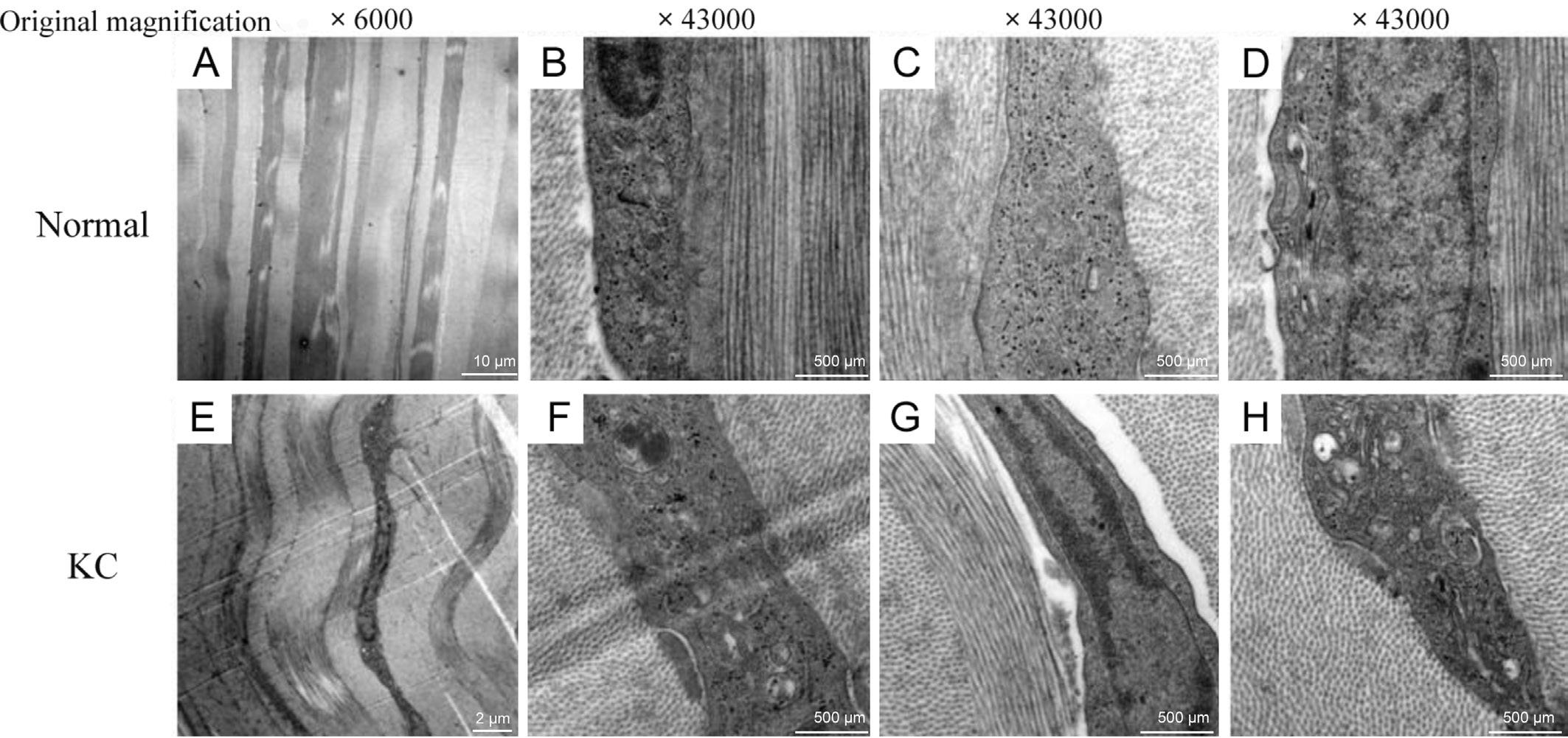
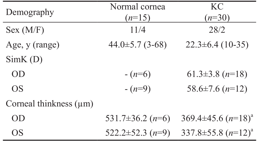
Figure 1 Ultramicroscopic structures of corneal stroma
A-D: Normal corneal stroma; E-H: KC stroma.
Western BlottingCellular total proteins were extracted according to the manufacturer's protocol by One Step Animal Tissue Active Protein Extraction Kit (Sangon Biotech, Shanghai,China). The protein concentration was determined by using a Modified Bradford Protein Assay Kit (Sangon Biotech,Shanghai, China). Equal amounts of protein (15 μg) were loaded and separated by 12% SDS-PAGE gel and then transferred onto a PVDF membrane. The membrane was soaked with 5% milk for 2h. After washing with PBSTween-20 (PBST; 5min/wash), the membrane was incubated with the primary antibodies including VSX1 (1:500, Santa Cruz Biotechnology, Inc., California, USA), α-SMA (1:1000,Sigma-Aldrich, Inc., St. Louis, Mo, USA) and GAPDH(1:1000, Beijing Biosynthesis Biotechnology Co., Ltd.,Beijing, China) at 4℃ overnight. The membranes then were washed with PBST three times and incubated at room temperature for 2h with the HRP-conjugated secondary antibody (1:5000, Thermofisher Scientific,Massachusetts,USA). After that, it was washed three times with PBST and detected using the High sensitive ECL luminescence reagent(Sangon Biotech, Shanghai, China). Then, the relative band density was quantified using odyssey FC 5.25 (LI-COR Biotechnology, Nebraska, USA). GAPDH was used as the internal control.
Statistical AnalysisThe data were expressed as the mean±standard error of the mean. Independent-samples t-test was used for comparing the differences between normal and KC groups. IBM SPSS Statistics 19 was used for statistical analysis. P<0.05 was considered to indicate a statistically significant difference.
Ultramicroscopic StructureThe ultramicroscopic structure of normal and KC stroma were observed by SEM. Compared with normal corneal stroma, KC stroma showed several distinguishing properties. Fifirst of all, the collagen fibers were distortional and attenuate (Figure 1A, 1E). At the same time,the nucleus gap widened. Additionally, mitochondrion swells and mitochondrial cristae disappear. Golgi apparatus presents significant expansion and endoplasmic reticulum displays severe swelling (Figure 1B-1D, 1F-1H).
Expression of VSX1 in Keratoconus Corneas Immunoflurescence showed that VSX1 signal was clearly localized in the stroma of normal and KC cornea. Compared with normal tissue, increased VSX1 staining was detected in the KC (Figure 2A). These data were confirmed by RT-qPCR and Western blot respectively. Results in Figure 2B demonstrated that VSX1 mRNA level in KC was about 3 times higher than normal corneal tissues (P<0.001). Western blotting confirmed significantly VSX1 protein up-regulation in KC tissues (Figure 2C, P<0.001). Together, these results demonstrate VSX1 up-expression in KC tissues.
Expression of α-SMA in Keratoconus Corneas Immuno fl urescence showed that α-SMA also localised within the stroma. There is almost no immunostaining was observed in normal cornea, however, for KC, stronger immunostaining of α-SMA compared to normal cornea (Figure 3A). RT-qPCR assay results in Figure 3B showed that α-SMA mRNA level was about 1.5 times higher in the KC tissues than that in normal cornea tissues (Figure 3B; P<0.0001). Western blotting also showed that α-SMA protein expression was increased in KC tissues (Figure 3C; P=0.0002).
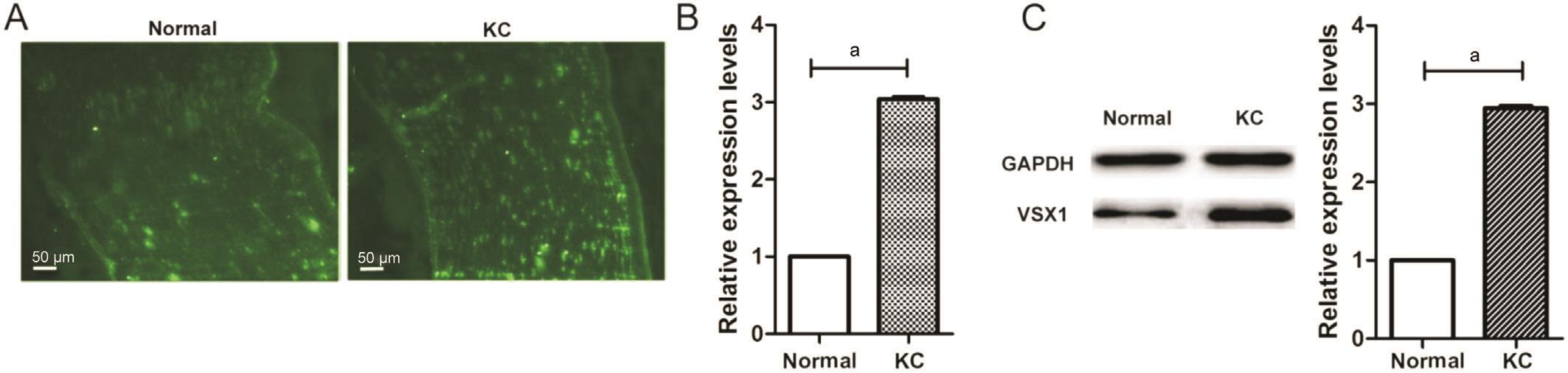
Figure 2 VSX1 up-expression in KC tissues compared to normal cornea tissues
A: Immuno fl urescence showed the expression level of VSX1. Scale bars: 50 µm; B: RT-qPCR analysis of VSX1 mRNA expression levels; C: Western blot analysis of VSX1 protein expression levels.aP<0.0001, independent-samples t-test.
Figure 3 α-SMA up-expression in KC tissues compared to normal cornea tissues
A: Immuno fl urescence showed the expression level of VSX1; B: RT-qPCR analysis of α-SMA mRNA expression levels; C: Western blot analysis of α-SMA protein expression levels. aP<0.001,independent-samples t-test.
KC is a corneal thinning disease whose etiology remains unknown. The pathophysiology of KC is characterized by the presence of a thin corneal stroma, altered extracellular matrix assembly and myo fibroblast differentiation[16]. The etiology of KC, which is multifactorial with genetic and environmental influences, remains elusive[17]. While many studies examine how environmental factors influence disease development,finding that the genetic triggers has been a major emphasis of KC research[18]. A large number of candidate genes have been studied in relation to KC pathogenesis. The link between VSX1 variants and KC susceptibility was first reported in Canada by Heon et al[19] and then many other studies had examined the potential mutations of VSX1 in KC patients. It is reported that the susceptibility of mutations in VSX1 can vary in different ethnic groups[6-7,20-28]. Our team also reported 3 SNPs(rs56157240, rs12480307 and rs6050307) in VSX1 associated with the susceptibility of Chinese KC patients, however,the actual function of VSX1 is unclear[10]. As a fifirst step to undefirstand the possible role of VSX1, we examined patterns of VSX1 expression in both human normal and KC. Different from previous research that VSX1 was not detected in mice cornea[29], in our study, VSX1 was expressed slightly in the stromal of normal human corneas but strongly in KC.
The loss of structural integrity in the KC was caused by the presence of abnormal keratocytes and matrix proteins.Keratocytes are activated into myo fibroblast, causing abnormal secretion of extracellular matrix[3]. Our SEM results confirmed that the collagen fibers of KC stromal were distortional and attenuate and keratocytes were abnormal changed, revealing that the reduction of the interfibrillar distance of collagen sheets and the increase of proteoglycans with abnormalities in their configuration as the condition evolves. Barbaro et al[30]had analyzed VSX1 expression and its involvements in wound-healing processes, in which differentiation of corneal keratocytes into myofibroblasts occurs and myofibroblastspecific marker (α-SMA) expresses. They considered that VSX1 sharply high expressed in the corneal stroma may elucidate the pathophysiological origin of KC. Xu et al[31]investigated the effects of bovine pituitary extract on the proliferation of keratocytes and confirmed that the expression of VSX1 reached a high level when many keratocytes underwent fibroblastic transformation. In this study, we also have analyzed α-SMA expression in normal human and KC,which is absent in the stromal of normal human cornea but intense expression in KC. The results indicate that the upgrade expression of VSX1 might cause the intense expression of α-SMA in the keratocytes, which may lead to differentiation of corneal keratocytes into myofibroblasts. The presence of abnormal keratocytes could alter the secretion of extracellular matrix and the structure of corneal stroma,which is the causation of stroma thinning and keratoconus occurrence.
In summary, compared with normal corneal stroma, we found that the expression level of VSX1 and α-SMA in KC increased.Taken together previous SNP studies, these observations indicate that VSX1 is related to the activation of keratocytes and involved in the pathogenesis of keratoconus. However,it remains to be determined whether the increased expression of VSX1 observed is directly involved in KC pathogenesis or is secondary responses. The specific role of VSX1 in KC pathogenesis needs further study.
We are grateful to all subjects who kindly agreed to participate in this study. We also appreciate the support and cooperation of the staff of Xi'an Eye Bank.
Foundations:Supported by Natural Science Foundation of Shaanxi Province (No. 2017JM8040); Xi'an Science and Technology Project [No.2017116SF/YX010(7)].
Conflicts of Interest: Wang YN, None; Liu XN, None; Wang XD, None; Yin Y, None; Chen Y, None; Xiao XH, None; Xu K, None; Zhu XP, None.
1 Krachmer JH, Feder RS, Belin MW. Keratoconus and related noninflammatory corneal thinning disorders. Surv Ophthalmol 1984;28(4):293-322.
2 Barsam A, Petrushkin H, Brennan N, Bunce C, Xing W, Foot B, Tuft S. Acute corneal hydrops in keratoconus: a national prospective study of incidence and management. Eye (Lond) 2015;29(4):469-474.
3 Meek KM, Tuft SJ, Huang Y, Gill PS, Hayes S, Newton RH, Bron AJ.Changes in collagen orientation and distribution in keratoconus corneas.Invest Ophthalmol Vis Sci 2005;46(6):1948-1956.
4 Galvis V, Sherwin T, Tello A, Merayo J, Barrera R, Acera A. Keratoconus:an inflammatory disorder? Eye (Lond) 2015;29(7):843-859.
5 Bardak H, Gunay M, Yildiz E, Bardak Y, Gunay B, Ozbas H, Bagci O.Novel visual system homeobox 1 gene mutations in Turkish patients with keratoconus. Genet Mol Res 2016;15(4).
6 Guan T, Wang X, Zheng LB, Wu HJ, Yao YF. Analysis of the VSX1 gene in sporadic keratoconus patients from China. BMC Ophthalmol 2017;17(1):173.
7 Shetty R, Nuijts RM, Nanaiah SG, Anandula VR, Ghosh A, Jayadev C, Pahuja N, Kumaramanickavel G, Nallathambi J. Two novel missense substitutions in the VSX1 gene: clinical and genetic analysis of families with Keratoconus from India. BMC Med Genet 2015;16:33.
8 Moussa S, Grabner G, Ruckhofer J, Dietrich M, Reitsamer H. Genetics in keratoconus-what is new? Open Ophthalmol J 2017;11:201-210.
9 Vincent AL, Jordan C, Sheck L, Niederer R, Patel DV, McGhee CN.Screening the visual system homeobox 1 gene in keratoconus and posterior polymorphous dystrophy cohorts identifies a novel variant. Mol Vis 2013;19:852-860.
10 Wang Y, Jin T, Zhang X, Wei W, Cui Y, Geng T, Liu Q, Gao J, Liu M,Chen C, Zhang C, Zhu X. Common single nucleotide polymorphisms and keratoconus in the Han Chinese population. Ophthalmic Genet 2013;34(3):160-166.
11 Levine EM, Hitchcock PF, Glasgow E, Schechter N. Restricted expression of a new paired-class homeobox gene in normal and regenerating adult goldfish retina. J Comp Neurol 1994;348(4):596-606.
12 Semina EV, Mintz-Hittner HA, Murray JC. Isolation and characterization of a novel human paired-like homeodomain-containing transcription factor gene, VSX1, expressed in ocular tissues. Genomics 2000;63(2):289-293.
13 Valgaeren H, Koppen C, Van Camp G. A new perspective on the genetics of keratoconus: why have we not been more successful?Ophthalmic Genet 2018;39(2):158-174.
14 Belin MW, Duncan JK. Keratoconus: The ABCD Grading System.Klin Monbl Augenheilkd 2016;233(6):701-707.
15 Livak KJ, Schmittgen TD. Analysis of relative gene expression data using real-time quantitative PCR and the 2(-Delta Delta C(T)) Method.Methods 2001;25(4):402-408.
16 Gokul A, Vellara HR, Patel DV. Advanced anterior segment imaging in keratoconus: a review. Clin Exp Ophthalmol 2018;46(2):122-132.
17 Gordon-Shaag A, Millodot M, Shneor E, Liu Y. The genetic and environmental factors for keratoconus. Biomed Res Int 2015;2015:795738.
18 Karolak JA, Gajecka M. Genomic strategies to undefirstand causes of keratoconus. Mol Genet Genomics 2017;292(2):251-269.
19 Héon E, Greenberg A, Kopp KK, Rootman D, Vincent AL, Billingsley G, Priston M, Dorval KM, Chow RL, McInnes RR, Heathcote G, Westall C, Sutphin JE, Semina E, Bremner R, Stone EM. VSX1: a gene for posterior polymorphous dystrophy and keratoconus. Hum Mol Genet 2002;11(9):1029-1036.
20 Bisceglia L, Ciaschetti M, De Bonis P, Campo PA, Pizzicoli C, Scala C, Grifa M, Ciavarella P, Delle Noci N, Vaira F, Macaluso C, Zelante L. VSX1 mutational analysis in a series of Italian patients affected by keratoconus: detection of a novel mutation. Invest Ophthalmol Vis Sci 2005;46(1):39-45.
21 Dash DP, George S, O'Prey D, Burns D, Nabili S, Donnelly U, Hughes AE, Silvestri G, Jackson J, Frazer D, Héon E, Willoughby CE. Mutational screening of VSX1 in keratoconus patients from the European population.Eye (Lond) 2010;24(6):1085-1092.
22 De Bonis P, Laborante A, Pizzicoli C, Stallone R, Barbano R, Longo C, Mazzilli E, Zelante L, Bisceglia L.Mutational screening of VSX1,SPARC, SOD1, LOX, and TIMP3 in keratoconus. Mol Vis 2011;17:2482-2494.
23 Eran P, Almogit A, David Z, Wolf HR, Hana G, Yaniv B, Elon P, Isaac A. The D144E substitution in the VSX1 gene: a non-pathogenic variant or a disease causing mutation? Ophthalmic Genet 2008;29(2):53-59.
24 Mintz-Hittner HA, Semina EV, Frishman LJ, Prager TC, Murray JC.VSX1 (RINX) mutation with craniofacial anomalies, empty sella, corneal endothelial changes, and abnormal retinal and auditory bipolar cells.Ophthalmology 2004;111(4):828-836.
25 Mok JW, Baek SJ, Joo CK. VSX1 gene variants are associated with keratoconus in unrelated Korean patients. J Hum Genet 2008;53(9):842-849.
26 Paliwal P, Singh A, Tandon R, Titiyal JS, Sharma A. A novel VSX1 mutation identified in an individual with keratoconus in India. Mol Vis 2009;15:2475-2479.
27 Saee-Rad S, Hashemi H, Miraftab M, Noori-Daloii MR, Chaleshtori MH, Raoofian R, Jafari F, Greene W, Fakhraie G, Rezvan F, Heidari M. Mutation analysis of VSX1 and SOD1 in Iranian patients with keratoconus. Mol Vis 2011;17:3128-3136.
28 Hao XD, Chen P, Chen ZL, Li SX, Wang Y. Evaluating the association between keratoconus and reported genetic loci in a Han Chinese population. Ophthalmic Genet 2015;36(2):132-136.
29 Watson T, Chow RL. Absence of Vsx1 expression in the normal and damaged mouse cornea. Mol Vis 2011;17:737-744.
30 Barbaro V, Di Iorio E, Ferrari S, Bisceglia L, Ruzza A, De Luca M,Pellegrini G. Expression of VSX1 in human corneal keratocytes during differentiation into myo fibroblasts in response to wound healing. Invest Ophthalmol Vis Sci 2006;47(12):5243-5250.
31 Xu ZZ, Li ZJ, Du LX, Li J, Wang LY. Using bovine pituitary extract to increase proliferation of keratocytes and maintain their phenotype in vitro.Int J Ophthalmol 2013;6(6):758-765.