INTRODUCTION
G laucoma is a progressive neurodegenerative optic neuropathy that results in morphological changes of optic nerve head (ONH) and loss of functional visual field[1].The elevation of intraocular pressure (IOP) is the most important risk factor for open angle glaucoma (OAG) and the effect of IOP reduction has been previously studied well[2]. In some cases, however, the loss of visual field progresses even with the well-controlled IOP, which indicates that other factors may be associated in the advancement of the glaucoma[3].
It has been suggested that vascular perfusion may have a role to the ocular ischemia and the death of the optic nerve.As prelaminar portion of ONH is perfused by branches of the peripapillary choroid, the function of the choroid can be important in the pathogenesis of glaucoma. Since abnormal perfusion of peripapillary choroid might be involved in the pathogenesis of glaucoma, peripapillary choroidal thickness(PCT) could also be concerned. Post-mortem histological studies have reported about thinner choroid in glaucoma[4].In particular, worsened choroidal and systemic blood flow parameters have been studied and may have affected the advancement of the glaucoma[5]. Some studies have revealed a reduced choroidal thickness (CT) in OAG due to the loss of both choriocapillaries and large vessels[4-5].
It has been reported a thicker foveal choroid in angle-closure glaucoma[6], a thinner peripapillary and macular choroid in glaucoma patients[7], whereas in other study no correlation was found between CT and glaucoma[8-9]. Although enhanced depth imaging (EDI) enabled choroidal visualization, the choroidalscleral boundary was difficult to discern. However, sweptsource optical coherence tomography (SS-OCT) uses a long wavelength that enhances its penetration[10]. The relationship may exist between CT and glaucoma structural parameters,because ONH and choroid are supplied by common branch of blood.
The goal of our study is to investigate the association between CT and OAG comparing patients affected by preperimetric glaucoma (PPG) and normal subject using SS-OCT. We also conducted a study to analyze the influence of factors including IOP, gender, age, retinal nerve fiber layer thickness (RNFLT),axial length (AL), and mean deviation (MD) on CT.
SUBJECTS AND METHODS
Ethical Approval This is a retrospective, comparative study assessing patients from October 2016 to July 2017. This study adhered to the tenets of the Declaration of Helsinki for research and followed all guidelines for investigation in human subjects. Written informed consent was obtained from each patient and the study protocol and informed consent were reviewed and approved by the institutional review board (IRB)before initiation.
Totally 55 eyes of OAG, 40 eyes of PPG, and 40 eyes of agematched normal eyes were analyzed by medical records.A comprehensive ophthalmologic evaluation was done including standard automated perimetry (Humphrey Visual Field Analyzer; Carl Zeiss-Meditec Inc., Dublin, CA, USA),color disc photography, Goldmann applanation tonometry, SS OCT (DRI-OCT system; Topcon, Tokyo, Japan) and IOL Master(Carl Zeiss Meditec, Dublin, CA, USA), and gonioscopy.Experienced ophthalmologists performed SS-OCT.
We included patients with open angle on gonioscopy and excluded those with intraocular disease or neurologic disease.All analysis used SPSS software version 19.0 (SPSS, Inc.,Chicago, IL, USA). Normally distributed data were expressed as mean±standard deviation. Univariate and multivariate linear regressions were applied to evaluate the association between CT and age, AL, central corneal thickness (CCT), RNFLT,and visual field MD. For all analyses, the level of statistical significance was set at P<0.05.
RESULTS
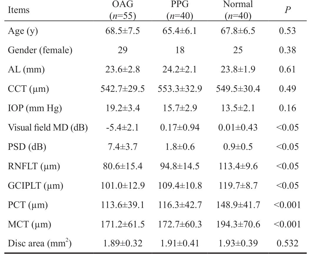
This study assessed 135 eyes, 55 eyes affected by OAG, 40 eyes of PPG, and 40 eyes of age-matched normal eyes. The demographic and ocular characteristics are summarized in Table 1. No significant difference was shown between groups in CCT, IOP, gender, and age (P>0.05).
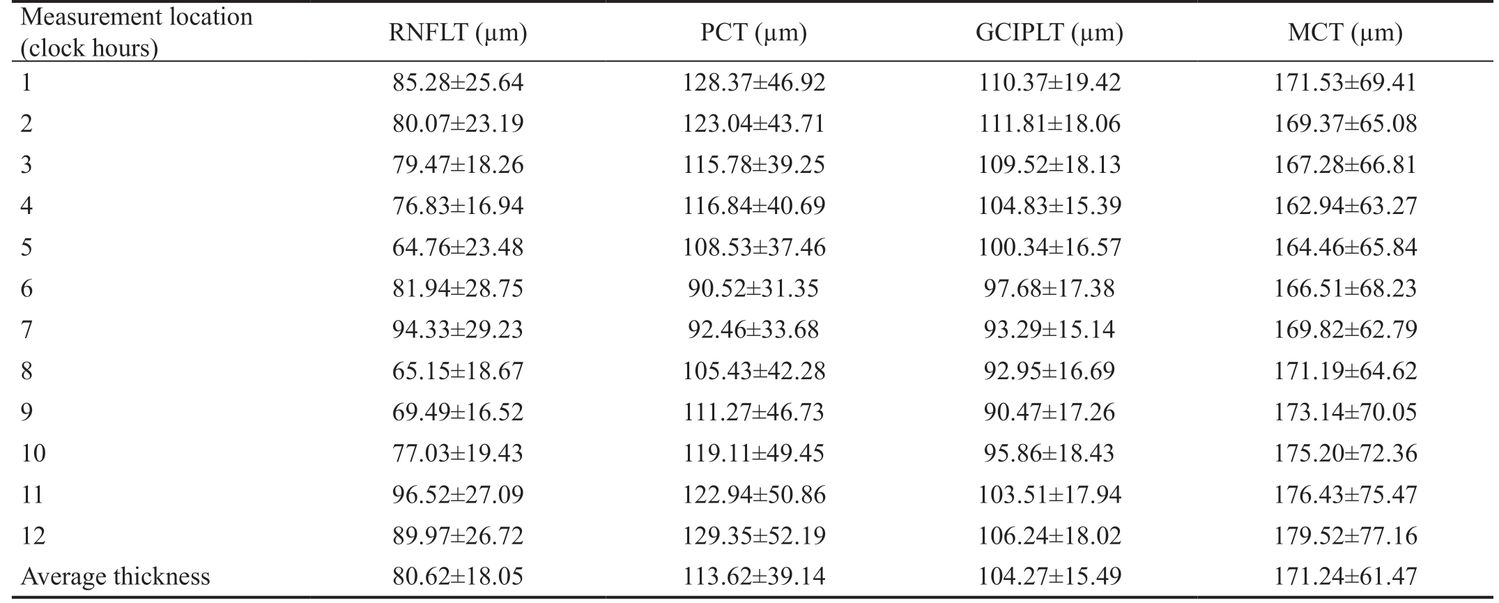
Mean RNFLT was 80.6±15.4, 94.8±14.5, 113.4±9.6 µm in OAG, PPG, and normal eyes, respectively (P<0.05). PCT was 113.6±39.1, 116.3±42.7, and 148.9±41.7 µm in OAG, PPG,and normal eyes, respectively (P<0.001). The difference in PCT was still left, after adjusting for age, AL and also after adjusting for age, AL, disc area (P=0.005). But the difference in macular CT (MCT) did not continue after adjusting for age,AL (P=0.084). No significant correlations were revealed with gender, CCT, or IOP (all P>0.05). The PCT showed significant differences between glaucoma and healthy eyes in 12 clockhour sectors and those continued after adjusting for age, AL and for age, AL, disc area. There was a statistically significant correlation between mean RNFLT and PCT in glaucomatous eyes. The 1, 2, 6, and 7 clock hours scans revealed a significant correlation (r=0.41, P=0.04; r=0.37, P=0.03; r=0.39, P=0.04;r=0.36, P=0.02, respectively; Table 2). CCT and IOP revealed
no significant difference between groups (P>0.05). In OAG group peripapillary choroid was thickest (129.35±52.19 µm) at the 12 clock hour and thinnest (90.52±31.35 µm) at the 6 clock hour, whereas RNFLT was thickest (96.52±27.09 µm) at the 11 clock hour and thinnest (64.76±23.48 µm) at the 5 clock hour(Table 2).
Mean ganglion cell-inner plexiform layer thickness (GCIPLT)was 101.0±12.9, 109.4±10.8, and 119.7±8.7 µm in OAG,PPG, and normal eyes, respectively (P<0.05). Mean MCT was 171.2±61.5, 172.7±60.3, and 194.3±70.6 µm in OAG, PPG,and normal eyes, respectively (P<0.001). The macular choroid was thinner in OAG and PPG group compared to normal eyes with statistically significant difference (P<0.001). The MCT showed differences among the groups, but they did not continue after adjusting for age, AL. No significant correlation was found between MCT and GCIPLT (P>0.05). There was no significant correlation between PCT and RNFLT or GCIPLT and MCT in normal eyes. Including AL, age, and gender thinner PCT was relevant with thinner RNFLT (β=0.438;F=5.47; P=0.03; R2=8%).
The association between CT in the peripapillary and macular area and variables including gender, AL, IOP, age, and CCT was evaluated. The peripaillary CT of OAG patients revealed a significant negative relation with AL (β=-7.04, P=0.027) in univariate regression and a significant relation with age (β=-0.97, P=0.004), AL (β=-9.49, P=0.001), and disc area (β=-8.36, P=0.035) in multivariate regression. The MCT of OAG
Table 1 Clinical characteristics of normal and glaucomatous patients

OAG: Open angle glaucoma; PPG: Preperimetric glaucoma; AL:Axial length; CCT: Central corneal thickness; IOP: Intraocular pressure; MD: Mean deviation; PSD: Pattern standard deviation;RNFLT: Retinal nerve fiber layer thickness; GCIPLT: Ganglion cell-inner plexiform layer thickness; PCT: Peripapillary choroidal thickness; MCT: Macular choroidal thickness.
Items OAG(n=55)PPG(n=40)Normal(n=40) P Age (y) 68.5±7.5 65.4±6.1 67.8±6.5 0.53 Gender (female) 29 18 25 0.38 AL (mm) 23.6±2.8 24.2±2.1 23.8±1.9 0.61 CCT (µm) 542.7±29.5 553.3±32.9 549.5±30.4 0.49 IOP (mm Hg) 19.2±3.4 15.7±2.9 13.5±2.1 0.16 Visual field MD (dB) -5.4±2.1 0.17±0.94 0.01±0.43 <0.05 PSD (dB) 7.4±3.7 1.8±0.6 0.9±0.5 <0.05 RNFLT (µm) 80.6±15.4 94.8±14.5 113.4±9.6 <0.05 GCIPLT (µm) 101.0±12.9 109.4±10.8 119.7±8.7 <0.05 PCT (µm) 113.6±39.1 116.3±42.7 148.9±41.7 <0.001 MCT (µm) 171.2±61.5 172.7±60.3 194.3±70.6 <0.001 Disc area (mm2) 1.89±0.32 1.91±0.41 1.93±0.39 0.532
patients revealed a significant association with AL (β=-15.39,P=0.008) in univariate regression and a relation with age(β=-2.03, P=0.003), AL (β=-20.94, P<0.001) in multivariate regression. No statistically significant correlations appeared with IOP, CCT, or MD (all P>0.05).
DISCUSSION
The loss of the choroidal vasculature was thought to be the cause of the decreased of CT. The thinning of choroid may be relevant with choroidal insufficiency which may result in the retinal ganglion cell damage of glaucoma. It has been suggested that the GCIPLT and RNFLT might be early structural markers when evaluating preperimetric and perimetric glaucoma.
Some previous OCT studies reported no difference in macular and PCT between OAG and healthy eyes[11-12]. However, some studies revealed thinning of PCT or MCT in OAG[13-14]. Hirooka et al[15] showed a relationship between CT and glaucoma severity especially. The results of some previous studies can't be believed since early glaucomatous optic disc change usually is focal. Since the choroidal vasculature supplies the anterior ONH, the peripapillary choroid may have a role in patients with glaucoma. Therefore, we supposed that PCT may be a practical method of showing the blood perfusion of ONH.However other study did not find an association between the PCT and glaucoma[16]. In our study, we discovered PCT thinning in OAG and PPG patients relative to normal controls.The PCT was related with disc area, AL, age but not with glaucoma severity (MD) in OAG. Hirooka et al[13] found that superior hemifield total deviation was worse than inferior which, indicates that PCT is thinner in areas of progressed glaucoma. In this study, PCT revealed no association with glaucoma severity in glaucomatous eyes. The reason of the difference could be explained by the fact that Hirooka et al[13]did not apply localized glaucoma severity parameter which we used in our study, and the PCT measurement point was different compared to our study. They also proposed that the area of the macular choroid closest to the ONH might be thinner in glaucoma without history of raised IOP. To evaluate this in our study, we investigated CT in the area between the macula and but found no association between CT and visual field MD.
Zhang et al[12] found that thinner CT was associated with older age and longer AL. Other studies reported that glaucomatous eyes showed thinner choroid. In this study, PCT revealed significant differences between glaucomatous and normal eyes and these differences remained after adjusting for AL, age, disc area. There are differences between previous studies and ours.First, the subject groups were different. Second, the manners of PCT measurement were different.
In present study, the results implied that CT was greatest at the macula and decreases along the peripapillary region which were consistent with previous studies but, CT was thinner in our study[17-18]. One possible reason is that previous reports used manual segmentation. Regarding MCT, there were significant differences between the groups, however these did not continue after adjusting for age and AL. These results are consistent with previous studies[19-20]. However, our results are different from previous studies at the other aspect. Mwanza et al[9] found out thicker MCT in OAG eyes than healthy eyes, although the result was not statistically significant. They supposed increased MCT was the result of a compensatory response of the choriocapillaries. However, that does not explain the study of Yin et al's[4], which reported reduced diameter of choroidal vessel.
Table 2 Evaluation of the RNFLT, peripapillary choroid, ganglion cell complex layer, macular choroid in OAG

RNFLT: Retinal nerve fiber layer thickness; OAG: Open angle glaucoma; PCT: Peripapillary choroidal thickness; GCIPLT: Ganglion cell-inner plexiform layer thickness; MCT: Macular choroidal thickness.
Measurement location(clock hours) RNFLT (µm) PCT (µm) GCIPLT (µm) MCT (µm)1 85.28±25.64 128.37±46.92 110.37±19.42 171.53±69.41 2 80.07±23.19 123.04±43.71 111.81±18.06 169.37±65.08 3 79.47±18.26 115.78±39.25 109.52±18.13 167.28±66.81 4 76.83±16.94 116.84±40.69 104.83±15.39 162.94±63.27 5 64.76±23.48 108.53±37.46 100.34±16.57 164.46±65.84 6 81.94±28.75 90.52±31.35 97.68±17.38 166.51±68.23 7 94.33±29.23 92.46±33.68 93.29±15.14 169.82±62.79 8 65.15±18.67 105.43±42.28 92.95±16.69 171.19±64.62 9 69.49±16.52 111.27±46.73 90.47±17.26 173.14±70.05 10 77.03±19.43 119.11±49.45 95.86±18.43 175.20±72.36 11 96.52±27.09 122.94±50.86 103.51±17.94 176.43±75.47 12 89.97±26.72 129.35±52.19 106.24±18.02 179.52±77.16 Average thickness 80.62±18.05 113.62±39.14 104.27±15.49 171.24±61.47
The current study has provided useful information about the association between glaucoma parameters and CT. In this study, a statistically significant correlation was shown between PCT and RNFLT of OAG eyes at 1, 2, 6, and 7 clock hours.It is important to remark that the correlation was highest at inferior and superior poles of the ONH which are known to be related to glaucomatous damage. In OAG eyes a significant correlation was reveled between PCT and RNFLT in OAG eyes.This study has some limitations. First, a relatively small number of eyes were evaluated. Second, our study was unable to assess the association between IOP-lowering eye drops and CT. CT might differ in untreated glaucoma patients. It is possible that elevated IOP may cause the reduction in CT secondary to IOP-induced reduction in choroidal blood flow.We're planning to study about it. Third, there is a possibility that PCT and MCT measurements did not acquire the exact same location in each subject. The main strength of our study is the utilization of available software, which permits automated segmentation and thickness measurements.
In conclusion, our study has shown that the PCT was decreased but did not reveal any relationship with glaucoma severity in glaucomatous eyes.
ACKNOWLEDGEMENTS
Foundation: Supported by the ICT R&D program of MSIT/IITP (No.2018-0-00242, Development of AI ophthalmologic diagnosis and smart treatment platform based on big data).
Conflicts of Interest: Park Y, None; Kim HK, None; Cho KJ, None.
1 Banitt M. The choroid in glaucoma. Curr Opin Ophthalmol 2013;24(2):125-129.
2 The Advanced Glaucoma Intervention Study (AGIS): 7. The relationship between control of intraocular pressure and visual field deterioration. The AGIS Investigators. Am J Ophthalmol 2000;130(4):429-440.
3 Wang W, Zhang XL. Choroidal thickness and primary open-angle glaucoma: a cross-sectional study and meta-analysis. Invest Ophthalmol Vis Sci 2014;55(9):6007-6014.
4 Yin ZQ, Vaegan, Millar TJ, Beaumont P, Sarks S. Widespread choroidal insufficiency in primary open-angle glaucoma. J Glaucoma 1997;6(1):23-32.5 Abegão Pinto L, Willekens K, Van Keer K, Shibesh A, Molenberghs G,Vandewalle E, Stalmans I. Ocular blood flow in glaucoma - the Leuven Eye Study. Acta Ophthalmol 2016;94(6):592-598.
6 Li Z, Wang W, Zhou M, Huang W, Chen S, Li X, Gao X, Wang J, Du S, Zhang X. Enhanced depth imaging-optical coherence tomography of the choroid in moderate and severe primary angle-closure glaucoma. Acta Ophthalmol 2015;93(5):e349-e355.
7 Dursun A, Ozec AV, Dogan O, Dursun FG, Toker MI, Topalkara A,Arici MK, Erdogan H. Evaluation of choroidal thickness in patients with pseudoexfoliation syndrome and pseudoexfoliation glaucoma. J Ophthalmol 2016;2016:3545180.
8 Hosseini H, Nilforushan N, Moghimi S, Bitrian E, Riddle J, Lee GY,Caprioli J, Nouri-Mahdavi K. Peripapillary and macular choroidal thickness in glaucoma. J Ophthalmic Vis Res 2014;9(2):154-161.
9 Mwanza JC, Hochberg JT, Banitt MR, Feuer WJ, Budenz DL. Lack of association between glaucoma and macular choroidal thickness measured with enhanced depth-imaging optical coherence tomography. Invest Ophthalmol Vis Sci 2011;52(6):3430-3435.
10 Yamanari M, Lim Y, Makita S, Yasuno Y. Visualization of phase retardation of deep posterior eye by polarization-sensitive swept-source optical coherence tomography with 1-microm probe. Opt Express 2009;17(15):12385-12396.
11 Zhang ZW, Yu MX, Wang F, Dai Y, Wu ZF. Choroidal thickness and open-angle glaucoma: a meta-analysis and systematic review. J Glaucoma 2016;25(5):e446-e454.
12 Zhang CW, Tatham AJ, Medeiros FA, Zangwill LM, Yang ZY, Weinreb RN. Assessment of choroidal thickness in healthy and glaucomatous eyes using swept source optical coherence tomography. PLoS One 2014;9(10):e109683.
13 Hirooka K, Tenkumo K, Fujiwara A, Baba T, Sato S, Shiraga F.Evaluation of peripapillary choroidal thickness in patients with normaltension glaucoma. BMC Ophthalmol 2012;12:29.
14 Roberts KF, Artes PH, O'Leary N, Reis AS, Sharpe GP, Hutchison DM, Chauhan BC, Nicolela MT. Peripapillary choroidal thickness in healthy controls and patients with focal, diffuse, and sclerotic glaucomatous optic disc damage. Arch Ophthalmol 2012;130(8):980-986.15 Hirooka K, Fujiwara A, Shiragami C, Baba T, Shiraga F. Relationship between progression of visual field damage and choroidal thickness in eyes with normal-tension glaucoma. Clin Exp Ophthalmol 2012;40(6):576-582.
16 Ehrlich JR, Peterson J, Parlitsis G, Kay KY, Kiss S, Radcliffe NM.Peripapillary choroidal thickness in glaucoma measured with optical coherence tomography. Exp Eye Res 2011;92(3):189-194.
17 Suh W, Cho HK, Kee C. Evaluation of peripapillary choroidal thickness in unilateral normal-tension glaucoma. Jpn J Ophthalmol 2014;58(1):62-67.
18 Margolis R, Spaide RF. A pilot study of enhanced depth imaging optical coherence tomography of the choroid in normal eyes. Am J Ophthalmol 2009;147(5):811-815.
19 Esmaeelpour M, Povazay B, Hermann B, Hofer B, Kajic V, Kapoor K, Sheen NJ, North RV, Drexler W. Three-dimensional 1060-nm OCT:choroidal thickness maps in normal subjects and improved posterior segment visualization in cataract patients. Invest Ophthalmol Vis Sci 2010;51(10):5260-5266.
20 Fujiwara T, Imamura Y, Margolis R, Slakter JS, Spaide RF. Enhanced depth imaging optical coherence tomography of the choroid in highly myopic eyes. Am J Ophthalmol 2009;148(3):445-450.