Dear Editor,
X EN Gel Stent 45 µm (Allergan Plc, Irvine, CA, USA)is a minimally-invasive procedure in glaucoma surgery perceived to reduce the risk of postoperative hypotony while sufficiently lowering the intraocular pressure (IOP)and requiring less topical antiglaucoma medications postoperatively.
Based on the Hagen-Poiseuille equation a micro-fistula's inner diameter of 45 µm and a total length of 6 mm may in theory create enough backpressure to prevent lowering of IOP below 6 mm Hg[1-2]. This case report highlights a rare but nonetheless important complication following this procedure.
CASE REPORT
A 76-year old woman with an 18-year history of pseudoexfoliative glaucoma in her right eye was referred for glaucoma surgery due to high IOP levels despite maximal topical therapy and temporary response to argon and selective laser trabeculoplasty. Topical therapy consisted of pilocarpine,timolol and bimatoprost while the patient was intolerant to brimonidine and dorzolamide. According to medical records the patient underwent uneventful cataract extraction with IOL implantation in the same eye 13 years ago.
Preoperative examination showed best-corrected visual acuity(BCVA) 20/20, IOP 30 mm Hg and glaucomatous optic disc excavation. The examination of the left eye did not show any signs of glaucoma. Subsequently, XEN45 was implanted uneventfully ab interno into the superonasal quadrant with 0.1 mL 0.02% mitomycin C (MMC) subconjunctivally.Routine postoperative topical steroids were prescribed.
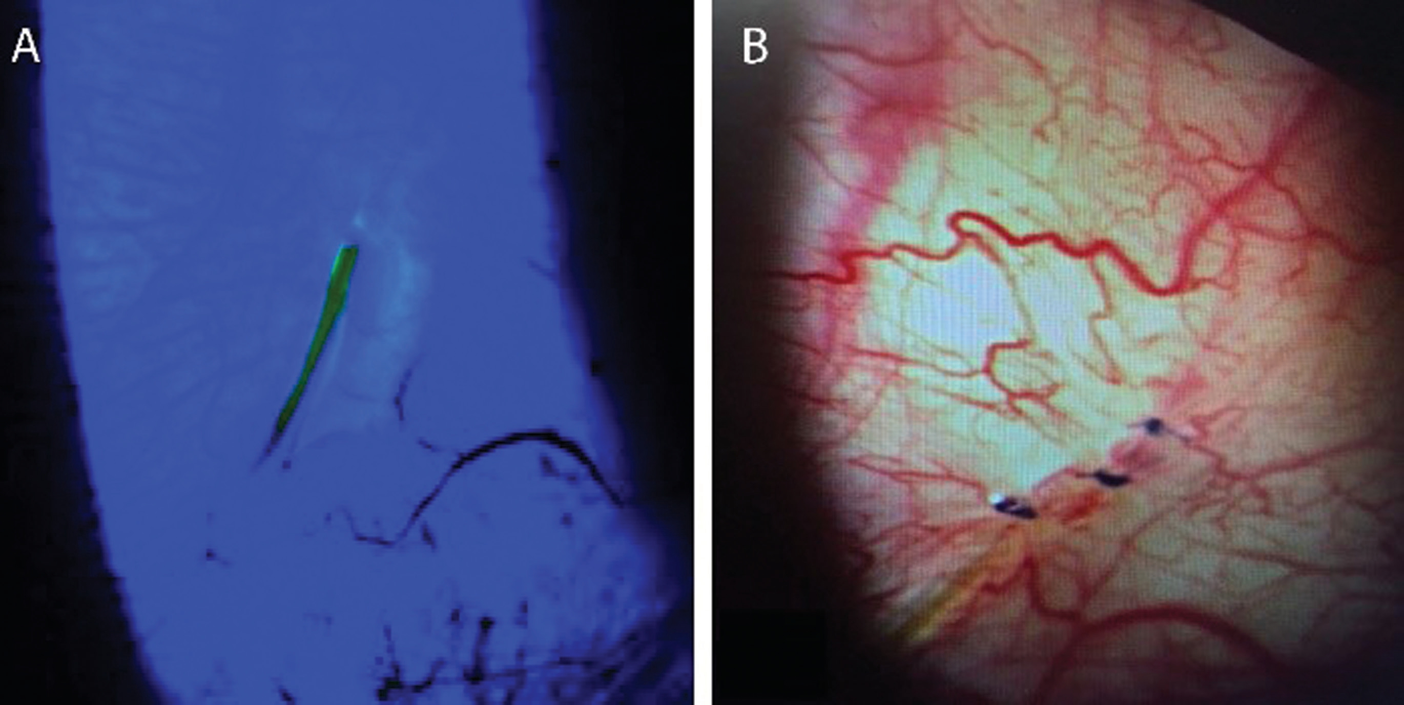
On first postoperative day, an IOP of 13 mm Hg was observed with an adequate filtrating bleb. However, the stent was slightly bent subconjunctivally. A pair of forceps were used to carefully straighten the implant without damaging overlying conjunctiva.Topical steroids were tapered within two months from surgery.The patient was then lost to follow-up and presented seven months later with BCVA 20/100, IOP 5 mm Hg and choroidal bullae. The distal microstent end had eroded overlying conjunctiva, exhibiting clear leakage which caused the low IOP (Figure 1A). Three single 9.0 Vicryl sutures were used to close the gap using the edges of the conjunctiva just above the stent (Figure 1B). Two days later IOP was still low (8 mm Hg).However, partial regress of choroidal bullae was noted.
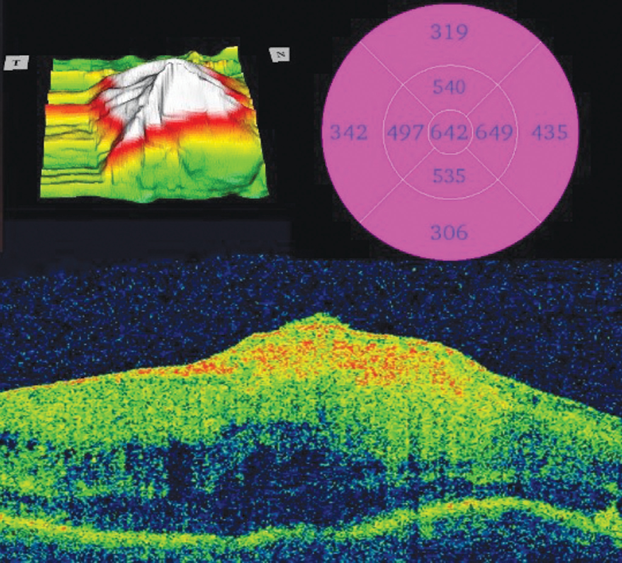
Unfortunately, the patient dropped out from follow-up,returning four months later due to worsening of visual acuity. Again, the distal part of the XEN stent was exposed,demonstrating local leakage causing low IOP (8 mm Hg).Until this time-point, no signs of subconjunctival scarring/bleb fibrosis had been observed nor ongoing active conjunctival inflammation. Spectral-domain optical coherence tomography(SD-OCT) confirmed a hypotony maculopathy (Figure 2).Consequently, the patient was brought to the operating theater where the exposed stent was covered by advancing healthy conjunctiva three to four millimeters anteriorly. Subsequently,topical steroids and NSAID were administered.
Within the following months the hypotony maculopathy persisted. The bleb surrounding the implant showed signs of increasing failure including scarring, a flat bleb appearance with dubious filtration and IOP increasing to 28 mm Hg while on maximal tolerable topical antiglaucoma medication.A clinical decision not to proceed with needling was made due to previous erosions, potential risk of new erosions with the use of 5-fluouracil, sensitive conjunctival tissue as well as low chance of succeeding since local tissue had already underwent multiple surgical manipulations. Thus, fornixbased trabeculectomy with 0.04% MMC was undertaken uneventfully. Postoperative period was subsequently uneventful and IOP remained ≤13 mm Hg without additional antiglaucoma therapy until last follow-up nine months later.
DISCUSSION
Through an ab interno approach inherently avoiding any incisional conjunctival manipulation the XEN Gel Stent is hypothesized to create less disruption to ocular surface anatomy, theoretically reducing the risk of subconjunctival fibrosis and as a consequence, risk of bleb failure. However,bypassing aqueous humor drainage through subconjunctival filtration creates a filtering bleb similar to trabeculectomy.The Hagen-Poiseuille equation states that by assuming a laminar flow, the outflow resistance of a non-compressible fluid through a cylindrical tube increases with increasing tube length as well as decreasing inner width. Thus, with a set length of 6 mm and an inner diameter of 45 µm, the XEN Gel implant is constructed to theoretically resist complications related to excessive filtration. Furthermore, studies have shown that the porcine gelatin material demonstrates excellent biocompatibility with ocular tissue, not causing any foreign-body reactions[1]. However, by nature of its construction the procedure does introduce foreign material into the ocular environment and even when properly located below conjunctiva, some modification to normal anatomy is inevitable. According to present studies episcleral fibrosis surrounding the implant does occur. As a consequence, bleb needling procedures have become accepted in a certain proportion of cases as part of postoperative care[3]. This case report highlights a potential risk for this new procedure such as exposure of the implant through the conjunctiva.

Figure 1 Color images of XEN45 Gel Stent exposure in ×10 magnification A: Complete transconjunctival stent erosion, only resistance to aqueous outflow remaining within the microstent. Slitlamp photograph showing fluorescein dye with cobalt blue filter.Aqueous leakage clearly visible on the right hand side of the stent. B:Following suturing of conjunctiva.

Figure 2 Hypotony maculopathy using SD-OCT Color-coded topographical maps (upper two maps) and horizontal B-scan (lower image) centered on the fovea.
XEN exposure was observed on two separate occasions completely eroding overlying conjunctiva. Interestingly, IOP never reached a level below 5 mm Hg despite the absence of conjunctival outflow resistance. Thus, XEN45 in this case appeared to provide enough aqueous outflow resistance to prevent hypotony below this level, illustrating in a real-life setting how the increasingly popular XEN45 microstent may constitute a hypotony-limiting factor in micro-invasive IOPlowering surgery.
A few possible risk factors for conjunctival erosion were identified. First, manipulation of conjunctiva took place prior to the exposures. Second, pseudoexfoliative glaucoma has been implicated with increased risk of erosion in a different, but not unrelated, setting of glaucoma drainage devices[4]. Although high implant flexibility and tissue conformation has been proposed to reduce risk of XEN erosion and migration, this might not be fully mitigated in the presence of non-autologous tissue[1]. Third, adjunctive MMC has been associated with wound leaks following trabeculectomy and could possibly be a contributing factor in the XEN Gel procedure as well[5]. Lastly,another potential risk factor for implant exposure might be the chronic use of multiple glaucoma eye drops causing a chronic local inflammation as well as thinning of the conjunctiva.
In summary, the ab interno microfistula created by the XEN Gel is not free of complications. Implant exposure, with potential risk of sight-threatening intraocular infections and hypotony-related complications are rare, but potential risks,that glaucoma specialists should be aware of. In our practice we have performed more than 130 cases since almost three years and this the only case of implant exposure, comparable to data from Schlenker et al[3].
ACKNOWLEDGEMENTS
Conflicts of Interest: Arnljots TS, None; Economou MA,
Receiving consulting fees from Allergan plc.
1 Lewis RA. Ab interno approach to the subconjunctival space using a collagen glaucoma stent. J Cataract Refract Surg 2014;40(8):1301-1306.2 Sheybani A, Reitsamer H, Ahmed II. Fluid dynamics of a novel microfistula implant for the surgical treatment of glaucoma. Invest Ophthalmol Vis Sci 2015;56(8):4789-4795.
3 Schlenker MB, Gulamhusein H, Conrad-Hengerer I, Somers A,Lenzhofer M, Stalmans I, Reitsamer H, Hengerer FH, Ahmed IIK.Efficacy, safety, and risk factors for failure of standalone ab internogelatin microstent implantation versus standalone trabeculectomy.Ophthalmology 2017;124(11):1579-1588.
4 Trubnik V, Zangalli C, Moster MR, Chia T, Ali M, Martinez P,Richman J, Myers JS. Evaluation of risk factors for glaucoma drainage device-related erosions: a retrospective case-control study. J Glaucoma 2015;24(7):498-502.
5 Palanca-Capistrano AM, Hall J, Cantor LB, Morgan L, Hoop J,WuDunn D. Long-term outcomes of intraoperative 5-fluorouracil versus intraoperative mitomycin C in primary trabeculectomy surgery.Ophthalmology 2009;116(2):185-190.