
Citation: Zhang QW, Zhai CB, Ma DL. Comparison of corneal curvature parameters obtained from two different instruments―Pentacam and VX120. Int J Ophthalmol 2019;12(8):1311-1316. DOI:10.18240/ijo.2019.08.12
・Clinical Research・
Comparison of corneal curvature parameters obtained from two different instruments―Pentacam and VX120
Qing-Wei Zhang, Chang-Bin Zhai, Dong-Li Ma
Beijing Institute of Ophthalmology; Beijing Tongren Eye Center; Beijing Key Laboratory of Ophthalmology and Visual Science, Beijing Tongren Hospital, Capital Medical University, Beijing 100730, China
Correspondence to: Qing-Wei Zhang. Beijing Institute of Ophthalmology; Beijing Tongren Eye Center; Beijing Key Laboratory of Ophthalmology and Visual Science, Beijing Tongren Hospital, Capital Medical University, Beijing 100730, China. zhaicbj@163.com
Received:
Abstract
AIM: To explore whether the same corneal curvature parameters and anterior chamber depth measured by Pentacam and VX120 have a good consistency and can replace each other.
METHODS: This study enrolled 140 eyes of 70 patients ranging in age from 19 to 53y. All eyes underwent a comprehensive ophthalmologic examination including an anterior segment analysis with the VX120 system (Visionix-Luneau Technologies, Chartres, France) and Pentacam (Oculus Optikgerate GmbH) respectively. The comparison on corneal curvature parameters was done between Pentacam and VX120 using clustered signed rank test; the interclass correlation coefficients (ICC) with 95% confidence intervals (CI) was calculated for each parameter between Pentacam and VX120; the Bland-Altman plot of each parameter was supplemented.
RESULTS: The anterior corneal curvature measured by VX120 was Ks: 44.00±1.78 D, KsAt: 89.45±22.18, Kf: 42.84±1.58 D, KfAt: 93.91±79.34; which measured by Pentacam was Ks: 43.80±1.82 D, KsAt: 91.17±21.40, Kf: 42.61±1.64 D, KfAt: 91.16±78.69. There was statistical difference between Pentacam and VX120 for anterior corneal curvature parameter (P<0.001). The posterior corneal curvature measured by VX120 was Ks: -6.42±1.23 D, KsAt: 91.00±23.45, Kf: -5.85±1.24 D, KfAt: 95.93±79.11; which measured by Pentacam was Ks: -6.44±0.32 D, KsAt: 92.24±11.75, Kf: -6.01±1.05 D, KfAt: 74.43±80.64. There was statistical difference between Pentacam and VX120 for posterior corneal curvature parameters (P<0.001). Anterior chamber depth (ACD) measured by Pentacam and VX120 was statistically different. Pentacam and VX120 achieved high consistency only on corneal anterior surface, including Ks and Kf. The ICCs were 0.96 (95%CI: 0.95, 0.97) and 0.95 (95%CI: 0.94, 0.97) respectively. For other corneal surface curvature parameters, all ICCs of between Pentacam and VX120 were below 0.87. Bland-Altman plots indicated of low consistency of corneal surface curvature parameters measured by Pentacam and VX120.
CONCLUSION: The corneal curvature parameters and anterior chamber depth measured by Pentacam and VX120 were statistically different. Data measured by Pentacam and VX120 is not suggested to replace each other, mixing data measured by Pentacam and VX120 together is not suggested either.
KEYWORDS: corneal curvature; anterior chamber depth; Pentacam; VX120; interclass correlation coefficients
DOI:10.18240/ijo.2019.08.12
Citation: Zhang QW, Zhai CB, Ma DL. Comparison of corneal curvature parameters obtained from two different instruments―Pentacam and VX120. Int J Ophthalmol 2019;12(8):1311-1316
INTRODUCTION
Measurement of the sharp, refractive power of the cornea are very important for refractive surgery design[1-3] and anterior segment diseases assessment[3]. Keratometry measures the corneal curvature and determines the corneal power and corneal astigmatism as well. A primary form of the keratometer was invented approximately 250 years ago[4]. Currently, a number of instruments are available for assessing corneal curvature, including Scheimpflug topography, optical low-coherence reflectometry, partial coherence interferometry and slit-scanning topography/pachymetry systems[5-9]. VX120 is a multi-diagnostic platform that commercially available recently, and has been shown to provide intersession consistent measurements of refraction and ocular aberrations[10], corneal curvature, eccentricity and aberrometric measurements[11] in health eyes. Given the optical principles behind the VX120 and Pentacam are same, the aim of the present study was to evaluate the agreement of the corneal curvature measurements and anterior chamber depth (ACD) obtained with the VX120 and another Scheimpflug-based topographic device⸺Pentacam.
SUBJECTS AND METHODS
Ethical Approval All procedures performed in studies involving human participants were in accordance with the ethical standards of the institutional and/or national research committee and with the 1964 Helsinki declaration and its later amendments or comparable ethical standards. Informed consent was obtained from all individual participants included in the study.
Seventy Chinese patients aged 19 to 53y with myopia or hyperopia who planned to received refractive surgery were included from the September 2014 to October 2016. A complete ocular examination was performed in all patients. Any ocular diseases other to refractive error were excluded.
All patients received examination of the anterior segment for both eyes. According to the random group, one group of patients received VX120 examination first, then Pentacam examination, and the other group received examination in the reverse order. Two observers took two instruments of each patient with each device. Each observer adjusted the keratometer eyepiece prior to use to avoid accommodative errors. In this study, the definition of ACD is distance from posterior point of corneal to anterior surface of ocular lens.
Pentacam Pentacam is a non-contact device which
uses combination of slit illumination system [that is light emitting diode
(LED) at 475 nm] and a rotating Scheimpflug camera to construct topographic images
of anterior chamber of eye, maps the shape and features of the corneal surface.
The simulated K readings (based on anterior corneal curvature alone) can be
obtained over a small central area (
VX120 System The VX120 system is a multi-diagnostic
platform that combines a Hartmann-Schack aberrometer, a Placido disk corneal
topographer, a Scheimpflug imaging-based system and an air tonometer. The
Hartmann-Shack aberrometer of the VX120 system measures 1500 points in 0.2s in
an area ranging from 2.0 to
Statistical
Analysis Mean and standardized deviation, as
well as median and interquartile, was used to make statistical description for
continuous variables according to the normality test results. Frequency and
proportions were used for statistical description of categorical variables.
Pearson correlation coefficients were used to evaluate the correlation of
different measurements done by Pentacam and VX120. The ICC was performed to
assess the consistency of different measurements done by Pentacam and VX120. Clustered signed rank test was used to make comparison between different
measurements, besides, due to that both eyes were included, a Rosner-Glynn-Lee
method was applied to make correction for the clustered data. The significance
level was set to be 0.05. The analysis was done using an open source R program
(version
RESULTS
A total of 70 subjects (140 eyes) was included in this study, and subjects’ average age was 29.69±7.32 years old, with male accounting for 30%. The median distant visual acuity of subjects was 0.08 (0.05, 0.15), the median near visual acuity was 1.00 (1.00, 1.00).
Correlation analysis of corneal surface curvature between Pentacam and VX120 was done (Table 1). Results showed that all corneal surface curvature parameters measured by Pentacam and VX120 were statistically correlated (P<0.001).
Table 1 Correlation analysis of corneal surface curvature between Pentacam and VX120
Variables |
r |
P |
Ks-anterior corneal curvature |
0.967 |
<0.001 |
Kf-anterior corneal curvature |
0.965 |
<0.001 |
Ks-posterior corneal curvature |
0.745 |
<0.001 |
Kf-posterior corneal curvature |
0.763 |
<0.001 |
BFS-anterior surface elevation |
0.887 |
<0.001 |
BFS-posterior surface elevation |
0.726 |
<0.001 |
Average anterior corneal curvature |
0.963 |
<0.001 |
Average posterior surface elevation |
0.756 |
<0.001 |
ACD |
0.599 |
<0.001 |
BFS: Best simulate sphere; ACD: Anterior chamber depth.
Comparison
on measurement of anterior and posterior corneal surface curvature parameters
between Pentacam and VX120 was further done (Table 2). For corneal anterior
surface, mean Ks obtained by Pentacam and VX120 was 43.80±1.82 D and 44.00±1.78
D respectively, the difference was statistically significant (Z=-4.022, P<0.001).
Mean Kf obtained by Pentacam and VX120 was 42.61±1.64 D and 42.84±1.58 D
respectively, the difference was statistically significant (Z=-5.189, P<0.001).
The same result was expressed on the posterior corneal surface measurement,
mean Ks was -6.44±0.32 D and -6.42±1.23 D for Pentacam and VX120 respectively,
the difference was statistically significant (Z=2.882, P=0.003),
and mean Kf was -6.01±1.05 D and -5.85±1.24 D for Pentacam and VX120
respectively, the difference was statistically significant (Z=-5.578, P<0.001).
There was statistical difference in the average anterior corneal curvature (Z=-4.566, P<0.001) and average posterior surface elevation either (Z=-4.172, P<0.001). There was no significant statistical difference in anterior
and posterior corneal meridian data between two devices. Moreover, we present
the best simulate sphere (BFS)-anterior surface elevation and BFS-posterior
surface elevation as well, statistical difference was observed between Pentacam
and VX120 (P<0.001). ACD measured by the VX120 and Pentacam was 3.15±
Table 2 Comparison on measurement of corneal surface curvature between Pentacam and VX120
Variables |
Pentacam |
VX120 |
Z |
P |
Ks-anterior corneal curvature |
43.80±1.82 |
44.00±1.78 |
-4.022 |
<0.001 |
KsAt-anterior corneal curvature |
91.17±21.40 |
89.45±22.18 |
-0.945 |
0.344 |
Kf-anterior corneal curvature |
42.61±1.64 |
42.84±1.58 |
-5.189 |
<0.001 |
KfAt-anterior corneal curvature |
91.16±78.69 |
93.91±79.34 |
-1.431 |
0.152 |
Ks-posterior corneal curvature |
-6.44±0.32 |
-6.42±1.23 |
2.882 |
0.003 |
KsAt- posterior corneal curvature |
92.24±11.75 |
91.00±23.45 |
1.271 |
0.203 |
Kf-posterior corneal curvature |
-6.01±1.05 |
-5.85±1.24 |
-5.578 |
<0.001 |
KfAt- posterior corneal curvature |
74.43±80.64 |
95.93±79.11 |
-1.870 |
0.061 |
BFS-anterior surface elevation |
7.90±0.27 |
7.86±0.29 |
4.940 |
<0.001 |
BFS-posterior surface elevation |
6.46±0.28 |
6.57±0.27 |
-6.717 |
<0.001 |
ACD |
3.15±0.29 |
2.89±0.23 |
6.609 |
<0.001 |
Average anterior corneal curvature |
43.2±1.69 |
43.42±1.63 |
-4.566 |
<0.001 |
Average posterior surface elevation |
-6.27±0.29 |
-6.24±0.78 |
-4.172 |
<0.001 |
ACD: Anterior chamber depth.
The ICCs of corneal surface curvature between Pentacam and VX120 were calculated furtherly (Table 3). Results showed that Pentacam and VX120 achieved high consistency only on corneal anterior surface, including Ks and Kf, the ICCs were 0.96 (95%CI: 0.95, 0.97) and 0.95 (95%CI: 0.94, 0.97) respectively. For other corneal surface curvature parameters, all ICCs of between Pentacam and VX120 were below 0.87, indicating of low consistency.
Table 3 Inter-class coefficients of corneal surface curvature between Pentacam and VX120
Variables |
ICC |
95%CI |
Ks-anterior corneal curvature |
0.96 |
0.95, 0.97 |
KsAt-anterior corneal curvature |
0.34 |
0.19, 0.48 |
Kf-anterior corneal curvature |
0.95 |
0.94, 0.97 |
KfAt-anterior corneal curvature |
0.46 |
0.32, 0.58 |
Ks-posterior corneal curvature |
0.15 |
0.02, 0.28 |
KsAt- posterior corneal curvature |
0.25 |
0.09, 0.40 |
Kf-posterior corneal curvature |
0.26 |
0.10, 0.41 |
KfAt-posterior corneal curvature |
0.10 |
0.01, 0.19 |
BFS-anterior surface elevation |
0.87 |
0.83, 0.91 |
BFS-posterior surface elevation |
0.65 |
0.55, 0.74 |
ACD |
0.26 |
0.12, 0.40 |
ICC: Intra class correlation; ACD: Anterior chamber depth.
We furtherly assessed the consistency of corneal surface curvature parameters between Pentacam and VX120 using Bland-Altman plot, as shown in Figure 1. Results showed that in spite of the low differences (biases) between values, the 95%CI limits were fairly wide, with many data points outside the 95%CI, indicating of low consistency of corneal surface curvature parameters measured by Pentacam and VX120.
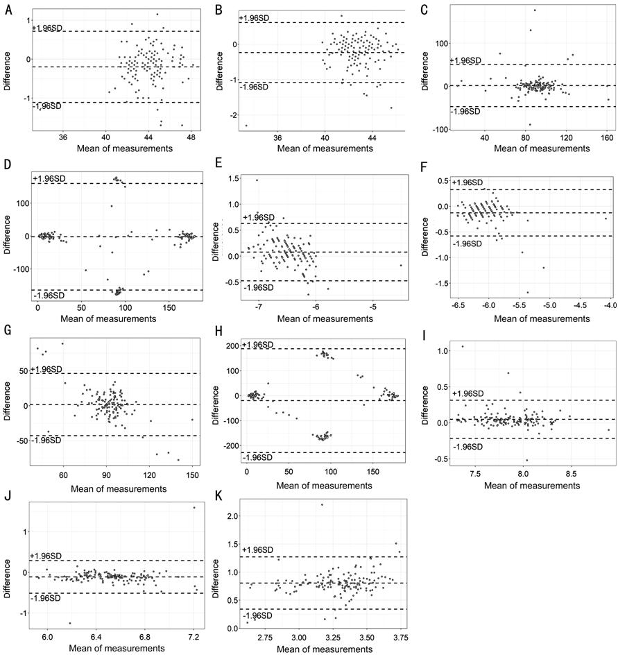
Figure 1 The consistency of corneal surface curvature parameters between Pentacam and VX120 using Bland-Altman plot A: Ks-anterior chamber curvature; B: Kf-anterior chamber curvature; C: KsAt-anterior chamber curvature; D: KfAt-anterior chamber curvature; E: Ks-posterior chamber curvature; F: Kf-posterior chamber curvature; G: KsAt-posterior chamber curvature; H: KfAt-posterior chamber curvature; I: BFS-anterior surface elevation; J: BFS-posterior surface elevation; K: Anterior chamber depth.
DISCUSSION
Corneal curvature measurement is essential to advances in refractive surgery and related fields. Successful outcomes require accurate & reliable data from the topography examination, inaccurate or unreliable data from the topography examination will lead to an inaccurate treatment.
Over the past few years, several instruments to image the anterior segment of the eye have been developed and made commercially available. Among the new technologies, optical coherence tomography, slit-scanning tomography and rotating Scheimpflug tomography currently play a major role[12].
We used to calculate corneal power using the anterior surface curvature multiplied by an index of refraction which assumes a fixed relationship between the anterior and posterior curvatures[13]. However, some studies indicated the inaccuracy in the default index of refraction and the corneal power is due to the non-persistent between anterior and posterior surface[14]. Scheimpflug-Placido based corneal tomographers are able to reconstruct three-dimensional images of the anterior segment and evaluate the whole cornea by obtaining information from both anterior and posterior corneal surfaces.
Recently, multifunctional diagnosis platform has become popular because they provide a “total workstation” concept. The validity of an instrument or procedure is generally expressed in terms of repeatability and agreement with another or with a standard reference[15]. Repeatability refers to the variation in measurements obtained by the same observer under same conditions over a short period of time. Agreement quantifies the similarity between any two measurements using different methods on the same subject. The limits of agreement, described by Bland and Altman[16], are defined as the mean difference ±1.96 D of differences. As mentioned before, VX120 has been demonstrated to have very good repeatability[10-11]. In this study, we undertook an initial evaluation of the VX120 Corneal Analysis System by comparing it to the Pentacam, which is the currently accepted standard for measuring the ocular anterior segment. The purpose of our study is to see whether the agreement between these devices is good enough so that the readings can be used interchangeably.
Oculus’ Pentacam combines a rotating Scheimpflug camera with a static camera to acquire multiple photographs of the anterior eye segment. The Scheimpflug camera rotates along with a monochromatic slit-light source around the optical axis to obtain the slit images. This rotating system performs a corneal scan from zero to 180° and each one of the photographs is an image of the cornea at a specific angle[17]. The static camera is placed in the centre to detect the pupil’s contours and control fixation.
Currently, there are some other commercially available instruments that are based on Scheimpflug imaging, such as the Galiei (Ziemer Ophthalmic Systems AG) the precision (Ligi Tecnologie Medicali) and the Sirius (CSO Ophthalmic). Although some scientific evidences confirming the consistency of the corneal measurements provided by different commercially available multi-diagnostic devices based on Scheimpflug imaging including Pentacam[18-21]. A Meta-analysis of agreement of various ophthalmic devices showed significant differences in mean posterior keratometry between Pentacam and Sirius. Significant difference in steep posterior keratometry was also noted between Pentacam and Galiei, only equivalent to Gailei for selected anterior and posterior keratometries (anterior steep keratometry, posterior: mean, steep and flat simulated keratometry)[22].
The results of this study showed that the mean curvature in the horizontal and vertical meridians differed significantly when measured by VX120 and Pentacam. VX120 providing a slightly steeper mean radius of curvature than the Pentacam. The same result was presented by L80, the predecessor of the VX120[23].
Anterior chamber depth is becoming a hot topic and plays an important role in correcting the refractive error after cataract surgery[24]. There is statistically significant difference on ACD measurement between VX120 and Pentacam. The value of VX120 is lower than Pentacam. This result seems conflict with the corneal curvature measurement.
Based on current result, we deduce that the corneal surface curvature and ACD measured by Pentacam and VX120 are different, using data measured by Pentacam to replace that measured by VX120 is not suggested. Mixing data measured by Pentacam and VX120 together is not suggested either. An offset incorporated into the instrument could mitigate the difference between the two instruments and make them interchangeable.
According to our knowledge, this is the first study to evaluate the agreement between VX120 and Pentacam, although the same mechanism the device based on. The data from our study showed they cannot use interchangeable. Just list the current Meta-analysis[25] showed, the agreement between the Scheimpflug imaging-based instruments is not satisfied, the measurement cannot be used interchangeably.
There are several shortages of our study: separate technician responsible for the different device, each measurement conducted only once due to the time constraints. We look forward to future studies that directly compare the performances of these devices.
ACKNOWLEDGEMENTS
We would like to thank Dr. Kai Cao for support of statistical analysis and Dr. Qing-Feng Liang for clinical support.
Foundation: Supported by the National Natural Science Foundation of China (No.81273806).
Conflicts of Interest: Zhang QW, None; Zhai CB, None; Ma DL, None.
REFERENCES